

The Science & Practice of Cryoneurolysis
Freeze the Pain
A comprehensive guide for patients and referring clinicians on one of the most exciting non-opioid pain technologies available today.
Chronic pain costs the United States more than $600 billion annually — and most of the burden is carried by the treatments we rely on most: opioids, repeated injections, and surgeries that never quite fix the underlying problem. Cryoneurolysis is not a cure-all, but it may be the most meaningfully different tool we’ve added to the interventional pain armamentarium in years.
THE BASICS
What Is Cryoneurolysis — and How Is It Different?
Cryoneurolysis uses precisely targeted, extreme cold to temporarily disable a specific peripheral nerve — interrupting its ability to transmit nerve signals (relieving pain and spasticity), without drugs, without burning tissue, and without permanent damage.
The technology most widely used in clinical practice today is the iovera° system (Pacira BioSciences), an FDA-cleared handheld device that delivers liquid nitrous oxide through a closed-end microneedle. This creates a focused ice ball at temperatures reaching -88°C at the tip — cold enough to cause a controlled, reversible nerve injury at the axon level, while leaving the structural nerve sheath (endoneurium, perineurium, epineurium) completely intact.
The result is a Sunderland Grade 2 axonotmesis — classically described as “nerve hibernation.” The nerve stops conducting pain immediately. Over weeks to months, the axon regenerates along its intact structural scaffold, restoring full nerve function. No permanent damage. No scar. No neuroma risk.
KEY DISTINCTION
Unlike radiofrequency ablation (RFA), which uses heat (85°C) and causes tissue damage, cryoneurolysis causes NO tissue damage. Unlike cryoablation used for tumors — which is clinically permanent at -140°C — cryoneurolysis is intentionally reversible. This reversibility is a feature, not a limitation.
Histological work by Hsu et al. (2014, Journal of Neurological Transmission) confirmed this using immunohistochemistry: at 2 weeks post-treatment, axonal architecture was disrupted; by 8 weeks, near-complete regeneration was visible — alongside a characteristic macrophage response consistent with Wallerian degeneration and nerve repair.
DURATION
How Long Does Relief Actually Last?
Clinical trial data — notably the Radnovich et al. 2017 randomized controlled trial in Osteoarthritis and Cartilage — demonstrates statistically significant, sustained pain relief out to 90 days. This is the FDA-cleared claim for iovera°.
In clinical practice, duration varies by anatomical target, proximity of treatment to the joint, and patient-specific biology. Outcomes frequently exceed the 90-day trial endpoint: 3–8 months in Knee Pain, 4–6+ months Shoulder Pain; 12–14 months Low Back Pain.
An important clinical insight: the more proximal the treatment point from the joint, the longer the nerve takes to regenerate to the target — effectively extending pain relief. Treating lumbar medial branches or knee genicular nerves farther from the articular surface can meaningfully prolong the duration of effect. This is something to thoughtfully individualize per patient.
For Meralgia Paresthetica (lateral femoral cutaneous nerve entrapment), relief duration in clinical experience can approach 1–2 years. Morton’s neuroma typically responds for 6–8 months per treatment cycle.
CLINICAL EVIDENCE
What Does the Research Show?
The evidence base for cryoneurolysis has grown substantially over the past decade, spanning joints, the spine, the chest wall, and the head. Below is a targeted summary of key studies informing current practice.
Knee Osteoarthritis
Radnovich et al. 2017 (Osteoarthritis and Cartilage) — the pivotal randomized, double-blind, sham-controlled trial of 180 patients — demonstrated statistically significant improvements in WOMAC outcomes at 6-month follow-up. Urban et al. 2021 (Arthroplasty Today, n=357) found no significant increase in postoperative infections when cryoneurolysis was performed two weeks preoperatively. Dasa et al. 2021 (Journal of Arthroplasty) found 45% less opioid usage in the treatment group at 6 and 12 weeks.
A 2024 real-world registry study in The Journal of Arthroplasty found that opioid-naïve patients receiving preoperative cryoneurolysis prior to TKA demonstrated improved pain scores, decreased opioid consumption, and meaningfully improved sleep disturbance over 6 months postoperatively.
Chronic Low Back Pain
A 2025 randomized pilot study in Pain Physician (30 patients) found that iovera° cryoneurolysis for facet-mediated chronic low back pain produced significantly lower pain scores at 180 days (3.1 vs. 5.4, p=0.01) compared to RFA, with functional disability also improving more substantially at one year. Functional outcomes on the Oswestry Disability Index were significantly lower with cryoneurolysis at 360 days.
Ankle Osteoarthritis
Perry et al. 2022 — a single-arm clinical trial of 40 patients with symptomatic ankle arthritis — demonstrated significant improvements in quality of life and pain scores following ultrasound-guided cryoneurolysis, an indication with historically limited non-surgical options.
Occipital Neuralgia
Kvarstein et al. 2019 (prospective multicenter, n=26): 70% of patients reported meaningful improvements and satisfaction at day 56. Grigsby et al. 2021 (double-blind randomized, n=52): greater than 50% improvement at 6–7 weeks in a controlled design.
Rib Fracture & Chest Wall Pain
Gabriel et al. 2020 — a sham-controlled RCT of 60 patients — demonstrated reduced opioid use and improved VAS scores. Multiple case series and pilot studies confirm cryoneurolysis’s role in reducing narcotic use in thoracic surgical and acute rib fracture contexts.
“Pain improvements can occur immediately and can last three months or more in the majority of cases — with meaningfully reduced opioid requirements in both surgical and non-surgical populations.”
PATIENT SELECTION
Who Is a Good Candidate?
Cryoneurolysis has one of the broadest applicability profiles of any interventional pain procedure I use. The right candidate is generally someone with a well-defined peripheral pain generator that can be accessed with a small-gauge needle under ultrasound guidance or anatomical localization. Conditions where I have observed particularly robust outcomes include knee arthritis (pre-operative, post-operative, and non-surgical), hip arthritis and labral pain via the PENG technique, shoulder arthritis and rotator cuff tendinopathy, ankle arthritis, Morton's neuroma, lumbar facet and sacroiliac joint pain, occipital neuralgia, cervicogenic headache, meralgia paresthetica, acute rib fracture, post-herpetic neuralgia, and spasticity-associated peripheral pain. The procedure is performed in a standard office or ambulatory surgery center setting under ultrasound guidance, takes minutes per target, and requires no sedation or fluoroscopy for most applications.
Absolute Contraindications
Cryoneurolysis should not be performed in patients with: open or infected wound at the treatment site, cryoglobulinemia, paroxysmal cold hemoglobinuria, cold urticaria, or Raynaud’s disease.
SAFETY PROFILE
What Are the Risks?
Cryoneurolysis has a favorable safety profile that compares well against other interventional options. Key considerations:
Dysesthesias — Rare, and typically only noticed after the local anesthetic wears off. Most discomfort represents the expected Wallerian degeneration process rather than a complication. This typically resolves.
Thermal skin injury — Rare and straightforward to manage. The iovera° device includes an integrated skin warmer that significantly mitigates this risk.
Transient muscle weakness — Reported in a minority of shoulder cases when the suprascapular nerve is treated within the notch. Duration is generally 2–3 weeks maximum.
Unlocking new pain generators — Occasionally, effective treatment of one nerve reveals an underlying pain source previously masked. This is not a complication; it is a diagnostic opportunity and should be communicated to patients proactively.
Histological studies confirm preservation of local arteries, veins, sebaceous glands, hair follicles, and skin cells. Any transient muscle injury resolves within 2–3 weeks. This is a meaningfully clean safety profile for an interventional procedure.
FOR REFERRING CLINICIANS
Cryoneurolysis is increasingly supported as a component of multimodal perioperative pain management and as a durable non-opioid option for chronic pain states where traditional approaches have provided inadequate relief. Appropriate referral candidates include patients with arthritis of the knee, hip, shoulder, or ankle who have failed conservative management; patients awaiting joint replacement with modifiable surgical risk factors; patients with neuralgia or nerve-mediated pain syndromes; and patients where opioid minimization is a priority. I welcome direct physician-to-physician consultation.
PERSPECTIVE
A Note on Where This Fits in Modern Pain Medicine
We are in an era defined by two competing realities: a chronic pain epidemic that affects more than 50 million Americans, and a long-overdue cultural reckoning with opioid dependency. Procedures like cryoneurolysis represent a third path — not a medication, not a surgery, not a corticosteroid injection cycling through diminishing returns — but a mechanistically different tool that respects the biology of pain and the reversibility patients deserve.
I am particularly interested in its application for active, middle-aged adults — the runners, lifters, tennis players, and weekend warriors who are not ready for joint replacement, who don’t want chronic opioids, and for whom a 3–12 month window of meaningful pain relief represents the difference between continued athletic participation and forced retirement from the activities that define their quality of life.
This is not a final answer for every patient. But for the right patient, at the right time, with the right target — it works remarkably well. And that is worth knowing about.
DISCLOSURE & REFERENCES
This article is for educational purposes and reflects clinical experience and interpretation of published literature. It is not a substitute for individualized medical evaluation. Key references: Radnovich et al. 2017 (Osteoarthritis Cartilage); Urban et al. 2021 (Arthroplasty Today); Dasa et al. 2021 (J Arthroplasty); McMillan et al. 2023 (Surg Technol Int); Gabriel et al. 2020 (RCT, rib); Perry et al. 2022 (ankle); Kvarstein et al. 2019 (occipital); Grigsby et al. 2021 (occipital RCT); Hsu et al. 2014 (J Neurol Trans); Ferillo ASRA 2024; Guynn et al. 2025 (Pain Physician).
ABOUT THE AUTHOR
Dr. Mahajer is a double board-certified physiatrist and sports medicine physician, fellowship-trained in Interventional Spine & Sports Medicine at the Icahn School of Medicine at Mount Sinai. He is an Assistant Professor of Neuroscience at FIU Herbert Wertheim College of Medicine. He is the Immediate Past President of the American Osteopathic College of Physical Medicine and Rehabilitation (AOCPMR), holds medical licenses in Florida, New York, and California, and has been recognized as a Top Physiatrist and Top Doctor in both Florida and New York.

Basivertebral Nerve Ablation for Chronic Low Back Pain
A Targeted, Thoughtful Approach to Lasting Relief
Chronic low back pain can be frustrating, exhausting, and life-limiting — especially when it persists despite physical therapy, medications, or injections. For some patients, the source of pain is not the muscles or discs alone, but the vertebral bones of the spine themselves. In these cases, basivertebral nerve ablation may offer a meaningful and lasting treatment option.
THE BASICS
What Is Basivertebral Nerve Ablation — and How Is It Different?
Not all low back pain comes from the same place. In a subset of patients, pain originates from within the vertebral bodies rather than from a pinched nerve or surrounding soft tissue. Small pain-sensing nerves embedded within the bone — the basivertebral nerves — detect these changes and generate a persistent, deep, aching pain that typically worsens with sitting, bending, or prolonged activity. On MRI, this pattern is often associated with Modic Type I or Type II endplate changes, signal alterations that reflect the underlying biology driving the pain. This type of pain does not respond reliably to injections targeting the facet joints or surrounding nerves, because the pain generator is not there. That is the clinical problem basivertebral nerve ablation is designed to solve.
Basivertebral nerve ablation is a minimally invasive outpatient procedure that uses controlled radiofrequency energy to interrupt the nerve signals responsible for this specific form of chronic low back pain. It is performed through a small access point in the skin using advanced imaging guidance, without placing any permanent hardware in the spine. The goal is not to mask pain but to address the underlying signal at its source.
KEY DISTINCTION
Unlike epidural steroid injections or medial branch blocks, which target the space around the spine or the facet joints, basivertebral nerve ablation targets the pain generator inside the vertebral body itself. Unlike spinal fusion, it involves no implants, no significant tissue disruption, and no prolonged recovery. It is not a replacement for surgery when surgery is truly indicated — it is a precision tool for a specific diagnosis that surgery was never well-suited to address in the first place.
FDA-cleared systems are available to perform this procedure, including the Boston Scientific Intracept® system and newer platforms such as the Stryker OptaBlate® BVN. The technology matters, but it is secondary to the most important variable in any interventional decision: patient selection.
DURATION
How Long Does Relief Actually Last?
The durability of basivertebral nerve ablation is one of its most clinically compelling features. The pivotal SMART trial and its long-term follow-up demonstrated sustained, statistically significant improvements in pain and function that have been maintained out to five years in treated patients. This is not a temporary block or a short-cycle injection — the ablation of the basivertebral nerve produces lasting interruption of the intraosseous pain signal, and because the nerve does not regenerate along the same pathway in the same way peripheral nerves do, the durability profile is fundamentally different from other ablative approaches in the spine.
CLINICAL EVIDENCE
What Does the Research Show?
The evidence base supporting basivertebral nerve ablation is among the strongest in the interventional spine space. The SMART trial — a randomized, double-blind, sham-controlled study — demonstrated statistically significant improvements in pain (VAS) and function (ODI) at six months, with the sham group subsequently crossing over to active treatment and showing comparable benefit. Long-term registry data from Fischgrund et al. and Becker et al. have confirmed durable outcomes at two and five years respectively, with clinically meaningful reductions in both pain scores and disability indices maintained across the follow-up period. Comparative effectiveness data suggest that BVNA outperforms standard care, including physical therapy and injections, in patients with confirmed Modic changes — and does so with a safety profile consistent with other minimally invasive outpatient spine procedures.
PATIENT SELECTION
Who Is a Good Candidate?
Patient selection is the most important determinant of outcome with basivertebral nerve ablation, and it is where I spend the most time in the evaluation process. The right candidate has had chronic low back pain for six months or longer, has not achieved lasting relief with conservative treatments including physical therapy and injections, has MRI findings consistent with Modic Type I or Type II endplate changes at one or more lumbar levels, and does not require urgent surgical intervention for instability, deformity, or neurological compromise. This is not a procedure I offer broadly. It is one I recommend when the diagnosis fits, the imaging supports it, and the patient's goals align with what the evidence shows this treatment can realistically deliver. A thorough clinical evaluation and careful imaging review are essential before any recommendation is made.
The procedure is performed on an outpatient basis under sedation. Most patients go home the same day and resume normal activities gradually under guidance. Because there are no implants and minimal tissue disruption, recovery is typically straightforward compared to surgical alternatives.
FOR REFERRING CLINICIANS
Basivertebral nerve ablation is increasingly recognized as an important option in the management of chronic axial low back pain driven by vertebral endplate pathology. Appropriate referral candidates include patients with chronic low back pain of six months or greater duration who have failed conservative management and have MRI findings consistent with Modic Type I or II changes; patients who have had an inadequate response to epidural or facet-based interventions; and patients for whom surgical options have been discussed but who prefer or require a non-implant, minimally invasive alternative. I welcome direct physician-to-physician consultation.
PERSPECTIVE
A Note on Where This Fits in Modern Spine Care
Chronic low back pain is the leading cause of disability in the United States, and the gap between how common it is and how well we treat it remains wide. For too long, the options have been framed as a binary — conservative care on one end, surgery on the other — with a middle ground defined largely by injections that were never designed to be curative. Basivertebral nerve ablation occupies a different position in that landscape. It is not a bridge to surgery. It is not a temporizing measure. For the right patient, it is a definitive treatment for a specific and underdiagnosed pain generator that has been responsible for years of suffering without a name.
I am particularly interested in identifying these patients early — before they have accumulated years of failed treatments, before their function has deteriorated significantly, and before surgery has become the only remaining option on the table. The evidence supports intervening once the diagnosis is confirmed and conservative care has been given a fair trial. Waiting longer does not improve outcomes. For the right patient, at the right time, basivertebral nerve ablation can be genuinely practice-changing. That is a meaningful thing to be able to offer.
DISCLOSURE & REFERENCES
This article is for educational purposes and reflects clinical experience and interpretation of published literature. It is not a substitute for individualized medical evaluation. Key references: Fischgrund et al. 2018 (SMART Trial, J Neurosurg Spine); Becker et al. 2021 (5-year outcomes, J Neurosurg Spine); Khalil et al. 2019 (Pain Med); Conger et al. 2021 (Pain Med); Friedly et al. 2021 (comparative effectiveness).
ABOUT THE AUTHOR
Dr. Mahajer is a double board-certified physiatrist and sports medicine physician, fellowship-trained in Interventional Spine & Sports Medicine at the Icahn School of Medicine at Mount Sinai. He is an Assistant Professor of Neuroscience at FIU Herbert Wertheim College of Medicine. He is the Immediate Past President of the American Osteopathic College of Physical Medicine and Rehabilitation (AOCPMR), holds medical licenses in Florida, New York, and California, and has been recognized as a Top Physiatrist and Top Doctor in both Florida and New York.
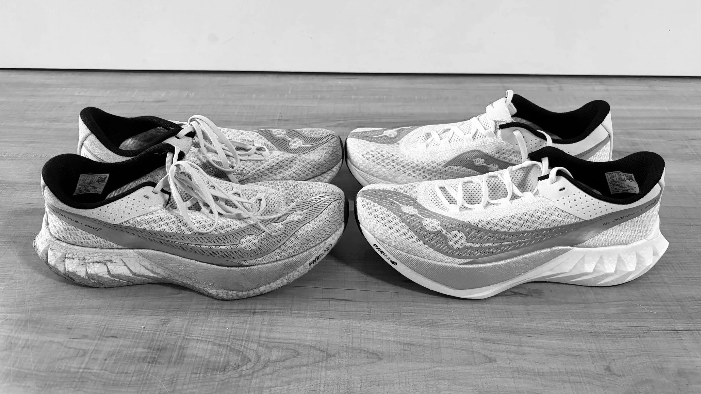
Starting to Run: What Your Body Actually Needs
The Right Running Shoes
As we step into a new year, many of us are thinking about improving our health, regaining consistency, and finally committing to the fitness goals we tend to push aside. The truth is simple: consistency and discipline outperform intensity every time. Small, repeatable actions stacked over time build stronger, healthier, more resilient bodies than short bursts of extreme effort ever will.
THE BASICS
Running as Medicine — and Why It Has to Be Approached Wisely
One of the most common New Year fitness goals is to begin or return to running. Whether the goal is to get back into shape, complete a first 5K, or train for a marathon, running remains one of the most effective and accessible ways to improve cardiovascular health, endurance, and mental well-being. There is a compelling physiological reason for this: training for endurance running has been shown to nearly double the density of mitochondria — the energy-producing organelles within muscle cells — improving cellular health and metabolic efficiency in ways that extend well beyond athletic performance. Running is powerful medicine. But like all powerful things, it needs to be approached wisely.
Great health requires multiple pillars: quality sleep, proper nutrition, meaningful social connection, mindfulness, and physical activity. When it comes to exercise specifically, two modalities are essential and complementary. Resistance training is critical for muscle preservation, metabolic health, joint protection, bone density, and longevity. Cardiovascular training is equally critical for heart health, endurance, brain function, energy efficiency, and disease prevention. We need both. Strength keeps us capable. Cardio keeps us thriving. Running conveniently addresses the cardiovascular requirement while providing weight-bearing benefits for bone and joint health when approached with appropriate progression.
STARTING RIGHT
Beginning — or Restarting — Running the Right Way
Most people who struggle with running or sustain early injuries do so not because their bodies cannot handle the activity, but because they progress too quickly. Modern life does not condition most of us for repetitive impact loading, and the body requires time to adapt its tendons, bones, and connective tissue even when cardiovascular fitness is already present. Your first weeks of running should feel easier than you expect. The goal is to build tolerance, not prove toughness.
Start with low weekly mileage and build gradually. Walk-run intervals are a smart and evidence-supported strategy, not a sign of weakness. Avoid chasing speed in the early weeks, and treat pain and soreness as information rather than a challenge to push through. The single most common cause of running injuries is increasing mileage, pace, or intensity too quickly — a pattern that leads predictably to overuse injuries including shin splints, plantar fasciitis, Achilles tendinopathy, anterior knee pain, and stress reactions in bone. A practical and well-supported guideline is to increase total weekly mileage by no more than 5 to 10 percent per week, with scheduled down weeks built into the training cycle.
EQUIPMENT
Your Feet Are Your Foundation
Footwear matters more than most runners appreciate, and the choices have never been more varied or more confusing. For the majority of runners, supportive footwear remains the appropriate starting point. The most important criterion is comfort — if a shoe does not feel right during the first mile, it is not the right shoe regardless of what the label says. Proper fit means adequate room in the toe box, a secure heel without slipping, and supportive midfoot contact without compression. Beyond fit, matching the shoe to the purpose matters: daily trainers for most runs, lightweight options for speed work once a base is established, cushioned shoes for longer distances and joint comfort, and trail-specific shoes for off-road terrain. Running shoes generally require replacement every 300 to 500 miles depending on body weight, running mechanics, and terrain — a variable most runners underestimate.
BUILDING DURABILITY
A Body That Loves to Run
Running places repetitive stress on muscles, tendons, ligaments, and bones. When introduced gradually, the body adapts exceptionally well to that stress — becoming stronger, more efficient, and more resilient over time. When introduced aggressively, it breaks down in predictable ways. The factors that determine which outcome you get are not complicated: gradual mileage progression, adequate rest and recovery between sessions, supplementary strength training to protect the joints and manage load, mobility work where individual limitations exist, quality sleep, and consistent nutrition and hydration. None of these are secrets. The challenge is executing them consistently rather than relying on motivation that tends to spike in January and erode by March.
FOR REFERRING CLINICIANS
Running-related injuries represent a significant and often undertreated source of musculoskeletal morbidity, particularly in the active adult population. Patients presenting with shin splints, plantar fasciitis, Achilles tendinopathy, patellofemoral pain, iliotibial band syndrome, or early stress reactions frequently benefit from a sports medicine evaluation that addresses both the injury and the underlying training error driving it. I offer comprehensive musculoskeletal assessment including diagnostic ultrasound, gait and biomechanical evaluation, and a full range of image-guided interventional options when appropriate — alongside evidence-based return-to-running programming. I welcome direct physician-to-physician consultation.
PERSPECTIVE
A Note on Consistency Over Intensity
We live in a culture that rewards extremes — extreme diets, extreme training programs, extreme transformation timelines. None of that is how healthy, durable fitness actually works. The athletes and active adults I see who sustain their fitness into their fifties, sixties, and beyond are not the ones who trained the hardest in their thirties. They are the ones who trained consistently, recovered intelligently, and respected the signals their bodies sent them before those signals became injuries. Running is one of the great democratizing forms of exercise — it requires no gym, no equipment beyond a decent pair of shoes, and no coach to begin. What it does require is patience and discipline applied over time. You do not need to be extreme this year. You need to be consistent. The body rewards that approach more reliably than any other.
DISCLOSURE & REFERENCES
This article is for educational purposes and reflects clinical experience and interpretation of published literature. It is not a substitute for individualized medical evaluation. Key references: Scharhag-Rosenberger et al. (running adaptation and mitochondrial biogenesis); Nielsen et al. 2014 (10% rule and injury prevention, BJSM); van Gent et al. 2007 (running injury incidence and risk factors, BJSM); Malisoux et al. 2015 (footwear and injury risk, AJSM).
ABOUT THE AUTHOR
Dr. Mahajer is a double board-certified physiatrist and sports medicine physician, fellowship-trained in Interventional Spine & Sports Medicine at the Icahn School of Medicine at Mount Sinai. He is an Assistant Professor of Neuroscience at FIU Herbert Wertheim College of Medicine. He is the Immediate Past President of the American Osteopathic College of Physical Medicine and Rehabilitation (AOCPMR), holds medical licenses in Florida, New York, and California, and has been recognized as a Top Physiatrist and Top Doctor in both Florida and New York.

What is Fusion?
Major Spine Surgery
Spinal fusion is a surgery designed to eliminate painful motion between two or more vertebrae by encouraging them to grow into a single, solid piece of bone. The plates, screws, rods, and cages used in these procedures are not the fusion itself — they are scaffolding that holds the spine steady while your body does the biological work of building new bone across the intended bridge. Understanding that distinction matters, because it changes how you think about the recovery, the risks, and the realistic expectations for what surgery can and cannot deliver.
THE BASICS
What Spinal Fusion Actually Does — and Why Mechanics Matter
Any surgery on the spine carries real short- and long-term consequences, and I think it is worth being direct about that. People often categorize procedures as minor or major, but any operation that alters spinal mechanics deserves serious consideration regardless of how it is labeled. By eliminating motion at one spinal level, fusion shifts mechanical stress to the segments immediately above and below. Over time, this additional workload can accelerate wear at those adjacent levels — a process called adjacent segment degeneration — and in some cases produce symptoms that require further treatment or revision surgery. Radiographic evidence of adjacent segment degeneration is commonly reported in the 20 to 40 percent range over time following lumbar fusion, with rates of revision surgery for symptomatic disease ranging from 7 to 20 percent depending on the study, the technique, and the length of follow-up. These are not reasons to avoid fusion when it is genuinely indicated. They are reasons to understand what you are agreeing to and to be appropriately selected before proceeding.
KEY DISTINCTION
The Complication Most Patients Do Not Hear Enough About
Pseudarthrosis — the failure of the intended fusion to fully consolidate — is one of the leading causes of ongoing pain after spinal fusion and a primary driver of what is commonly called failed back surgery syndrome. When the fusion does not take, persistent micro-motion remains at the intended fusion site, often producing pain that is indistinguishable from the original complaint and sometimes requiring revision surgery to address. The risk of pseudarthrosis increases meaningfully with the number of levels fused, and is compounded by smoking, older age, poorly controlled diabetes, osteoporosis, and suboptimal spinal alignment. Modern outcomes data suggest symptomatic pseudarthrosis occurs in approximately 2 to 3 percent of patients at ten years for single-level procedures, with risk rising substantially as more vertebral levels are included in the construct. This is not a rare or theoretical complication — it is a clinically important one that deserves an honest conversation before any patient proceeds to surgery.
CLINICAL EVIDENCE
What Does the Research Show?
The evidence on spinal fusion outcomes is nuanced and worth understanding in detail. Multilevel fusions carry more complications and produce less pain improvement on average than single-level procedures, a finding that has been replicated across multiple outcomes studies including Harada et al. 2021. In the cervical spine, contemporary data suggest that two-level fusions consolidate more reliably than three-level constructs, reinforcing the principle that biological and mechanical demands increase with each additional level added to a construct. Alignment matters significantly as well — malalignment following fusion is an established risk factor for both adjacent segment disease and pseudarthrosis, with one analysis estimating surgically relevant adjacent segment disease at approximately 2.4 percent per year following L4 to S1 fusion in the setting of poor alignment. Patients with poorly controlled diabetes face higher nonunion rates and worse overall outcomes, as documented by Steinmetz et al. in Spine Journal 2025. The literature on high-profile athletes, including Tiger Woods whose most recent lumbar surgery occurred in October 2025, illustrates how spinal mechanics and biology play out over years and across multiple procedures — a real-world pattern that reflects what the evidence predicts.
PATIENT SELECTION
When Fusion Is and Is Not the Right Answer
Surgery is never the first step in my practice, and fusion specifically is never a treatment for isolated axial low back pain without a clearly identified, surgically correctable structural problem. When I recommend surgical consultation, it is because conservative care has been exhausted and the pattern of pain correlates with a problem that surgery is genuinely well-suited to address, because progressive neurological deficit requires intervention to arrest nerve injury and prevent long-term disability, or because the patient is medically optimized and biologically positioned to heal. That last point is more important than most patients realize. A non-smoker with well-controlled metabolic health and adequate bone density has a fundamentally different risk profile than a patient who is actively smoking, has uncontrolled diabetes, or has significant osteoporosis. Addressing modifiable risk factors before proceeding to surgery is not a bureaucratic hurdle — it is how we improve the probability that the fusion actually works.
Patients with poorly controlled diabetes, active nicotine use, severe osteoporosis, or significant wound-healing risks carry higher complication and nonunion rates across the literature. I discuss these factors directly with every patient I evaluate for surgical referral, and I work with them to optimize whatever can be optimized before a recommendation is made.
FOR REFERRING CLINICIANS
Patients presenting for evaluation of potential spinal fusion benefit significantly from a thorough pre-surgical interventional medicine assessment — particularly to confirm the pain generator through diagnostic blocks, assess for non-surgical alternatives that may have been incompletely explored, and identify modifiable risk factors that could affect fusion outcomes. I offer comprehensive imaging review, diagnostic and therapeutic spinal injections, metabolic and musculoskeletal optimization guidance, and alignment-aware pre-surgical planning in collaboration with surgical partners. For patients who have undergone fusion and continue to experience pain, I provide post-surgical evaluation to distinguish adjacent segment pathology, pseudarthrosis, and other treatable causes from non-structural contributors to ongoing symptoms. I welcome direct physician-to-physician consultation.
PERSPECTIVE
A Note on Shared Decision-Making in Spine Care
Spinal fusion can be genuinely life-changing for the right problem, in the right patient, with meticulous planning and committed post-operative rehabilitation. It is not a cure for back pain broadly, and it is not a procedure whose consequences are limited to the operating room. The hardware is scaffolding. The fusion is biology. And biology does not always cooperate on the timeline or to the degree that either the patient or the surgeon hopes. What I can offer every patient who comes to me with this decision in front of them is a comprehensive evaluation, an honest interpretation of their imaging and clinical picture, and a clear-eyed discussion of all available options — from targeted injections and structured rehabilitation to surgical second opinions — so that whatever choice is made, it is made with full information and realistic expectations. That is the standard I hold myself to, and it is the only standard I think is acceptable when the stakes are this high.
DISCLOSURE & REFERENCES
This article is for educational purposes and reflects clinical experience and interpretation of published literature. It is not a substitute for individualized medical evaluation. Key references: Boonsirikamchai et al. 2024 (pseudarthrosis risk factors); Shahzad et al. 2023–2024 (symptomatic pseudarthrosis rates); Steinmetz et al. 2025 (diabetes and fusion outcomes, Spine J); Loggia et al. 2025 (alignment and adjacent segment disease, Spine J); Soh et al. 2025 (temporal patterns of ASD, J Clin Med); Okuda et al. 2018 (ASD after PLIF); Harada et al. 2021 (multilevel fusion outcomes); Nouh et al. 2012 (instrumentation principles).
ABOUT THE AUTHOR
Dr. Mahajer is a double board-certified physiatrist and sports medicine physician, fellowship-trained in Interventional Spine & Sports Medicine at the Icahn School of Medicine at Mount Sinai. He is an Assistant Professor of Neuroscience at FIU Herbert Wertheim College of Medicine. He is the Immediate Past President of the American Osteopathic College of Physical Medicine and Rehabilitation (AOCPMR), holds medical licenses in Florida, New York, and California, and has been recognized as a Top Physiatrist and Top Doctor in both Florida and New York.

Optimizing Knee Pain
Genicular Nerve Ablation
Chronic axial neck and back pain have long been the domain of radiofrequency ablation, and in my clinical practice I have seen countless patients benefit from this evidence-based technique. Over the past decade, the application of RFA has expanded meaningfully — not only for axial spine pain, but for chronic joint pain, with a growing and particularly compelling body of evidence centered on the knee.
THE BASICS
Genicular Nerve Radiofrequency Ablation — and Why Knee Pain Management Has Changed
The landscape of knee osteoarthritis treatment has shifted considerably in recent years, and for good reason. Corticosteroid injections, once a routine first-line option, are increasingly discouraged in many clinical contexts due to concerns over cartilage degradation and cumulative systemic effects. Hyaluronic acid injections have lost favor in multiple guidelines, offering limited long-term benefit for a significant proportion of patients. That has prompted a necessary shift toward recovery-oriented, rehabilitation-focused care — and it has created an opening for interventional approaches that address the neural drivers of pain rather than the joint environment alone. At Osso Health, I emphasize a multimodal, nonoperative approach to knee pain. I also recognize that for patients with end-stage osteoarthritis, total knee arthroplasty remains the definitive treatment. The clinical question I am most interested in is how we get patients there — and through it — in the best possible condition.
WHERE IT BEGAN
A Practice Built From the Hardest Cases First
My experience with genicular nerve ablation started where many pain stories end: after surgery. The first patients I treated with this technique were those experiencing persistent knee pain following total knee replacement — patients who had exhausted conservative options, were not candidates for revision surgery, and were living with ongoing pain that had no clear remaining treatment pathway. Using targeted thermal radiofrequency ablation of the genicular nerves, I was able to achieve meaningful pain relief and restore function for these patients without additional surgery or long-term medication dependence. Encouraged by those outcomes, I extended the same approach to patients with end-stage osteoarthritis who were delaying surgery for medical, logistical, or personal reasons. The results were consistent — offering a bridge that allowed them to maintain mobility, reduce medication reliance, and defer surgery while preserving quality of life.
KEY DISTINCTION
Thermal RFA, Cryoneurolysis, and How They Complement Each Other
Genicular nerve radiofrequency ablation uses controlled thermal energy delivered through image-guided needles to interrupt the sensory nerve pathways responsible for transmitting knee pain. It is performed under fluoroscopic or ultrasound guidance targeting the superolateral, superomedial, and inferomedial genicular nerves — the primary afferent contributors to knee joint pain. The procedure is outpatient, requires no implants, and produces no significant tissue damage beyond the targeted nerve. In addition to thermal ablation, I now offer cryoneurolysis for the genicular nerves — a technique that applies subzero temperatures to desensitize peripheral nerves through a reversible axonotmesis rather than thermal destruction. Cryoneurolysis offers a favorable sensory profile and may be particularly well-suited to patients with post-arthroplasty discomfort or those seeking temporary relief prior to planned surgery, where the reversibility of the effect is clinically advantageous.
CLINICAL EVIDENCE
What Does the Research Show?
The evidence base for genicular nerve RFA has grown substantially over the past decade. Multiple randomized controlled trials and systematic reviews have demonstrated statistically significant improvements in pain scores and functional outcomes compared to sham procedures and conservative care in patients with knee osteoarthritis. The preoperative application of genicular nerve RFA represents an emerging and particularly promising frontier. Early evidence supports this strategy, demonstrating improved postoperative pain control, enhanced early rehabilitation and mobilization, shorter hospital stays, fewer postoperative complications, and no increased risk of infection when RFA is performed prior to total knee arthroplasty. The mechanistic rationale is straightforward: by interrupting the chronic afferent pain signal before surgery, patients arrive in better neurological and functional condition for recovery, with lower baseline central sensitization and reduced perioperative opioid requirements.
PATIENT SELECTION
Who Is a Good Candidate?
Genicular nerve RFA is appropriate for patients with chronic knee pain secondary to osteoarthritis who have had an inadequate response to conservative management including physical therapy, oral medications, and intra-articular injections. It is also appropriate for patients with persistent pain following total knee arthroplasty who are not candidates for or do not wish to pursue revision surgery. Preoperative RFA should be considered for patients planning total knee arthroplasty who have significant chronic pain burden, high baseline opioid use, or risk factors for difficult postoperative pain management. As with all interventional procedures, precise patient selection and diagnostic accuracy are the primary determinants of outcome. A careful clinical evaluation and imaging review are essential before any recommendation is made.
FOR REFERRING CLINICIANS
Genicular nerve radiofrequency ablation and cryoneurolysis represent important additions to the perioperative and nonoperative management of knee pain, and I welcome referrals from orthopedic surgeons, primary care physicians, and other specialists managing this population. Whether the goal is optimizing a patient before planned total knee arthroplasty, managing persistent pain after joint replacement, or providing a durable nonoperative option for patients who are not surgical candidates, I offer a comprehensive evaluation, image-guided procedural expertise, and clear documentation back to the referring provider. My background includes extensive collaboration with surgeons across spine, total joint, upper extremity, and foot and ankle specialties. I understand the nuances of perioperative musculoskeletal care and the importance of a referring relationship built on communication and shared goals. I welcome direct physician-to-physician consultation.
PERSPECTIVE
A Note on Collaborative Perioperative Care
The management of chronic knee pain has for too long been treated as a binary — conservative care on one side, surgery on the other — with little attention paid to the interventional space between them and even less to what happens in the weeks and months surrounding the surgical episode itself. Perioperative pain management is not the surgeon's problem alone, and it is not solved by a standard anesthesia protocol. It requires a physician who understands the neuroscience of chronic pain, the biology of surgical recovery, and the specific nerve anatomy driving a given patient's experience. That is the role I aim to fill. When a patient arrives at surgery with years of central sensitization and a high baseline pain burden, the recovery is harder, the rehabilitation is slower, and the outcomes are less predictable. When that same patient has had their peripheral pain signal meaningfully reduced before the procedure, the entire postoperative course changes. That is not a theoretical benefit — it is what the evidence shows, and it is what I see in clinical practice. The opportunity to contribute meaningfully to surgical outcomes without being in the operating room is one I take seriously.
DISCLOSURE & REFERENCES
This article is for educational purposes and reflects clinical experience and interpretation of published literature. It is not a substitute for individualized medical evaluation. Key references: Choi WJ et al. 2011 (genicular nerve RFA, Pain); Ikeuchi M et al. 2011 (genicular nerve block and ablation outcomes); McCormick ZL et al. 2017 (systematic review, genicular RFA, Pain Med); Fonkoué L et al. 2019 (genicular nerve anatomy); Dasa V et al. 2021 (preoperative cryoneurolysis and TKA outcomes, J Arthroplasty); Radnovich R et al. 2017 (cryoneurolysis RCT, Osteoarthritis Cartilage).
ABOUT THE AUTHOR
Dr. Mahajer is a double board-certified physiatrist and sports medicine physician, fellowship-trained in Interventional Spine & Sports Medicine at the Icahn School of Medicine at Mount Sinai. He is an Assistant Professor of Neuroscience at FIU Herbert Wertheim College of Medicine. He is the Immediate Past President of the American Osteopathic College of Physical Medicine and Rehabilitation (AOCPMR), holds medical licenses in Florida, New York, and California, and has been recognized as a Top Physiatrist and Top Doctor in both Florida and New York.

Restoring Disc Health with Regenerative Medicine
Spine PRP
Disc-mediated low back pain is one of the most common and most undertreated conditions in spine medicine. For patients who have exhausted conservative care and are not ready for — or interested in — surgery, the question becomes what comes next. Intradiscal regenerative medicine — including platelet-rich plasma and, in select cases, bone marrow aspirate concentrate — represents one of the most promising answers currently available, and when performed with the right preparation, the right technique, and the right patient selection, the evidence supports its ability to provide meaningful and durable relief.
THE BASICS
What Is Intradiscal PRP and How Does It Work?
Platelet-rich plasma is derived from your own blood, processed to separate and concentrate the platelets that carry the growth factors responsible for initiating tissue repair. In the intradiscal application, this concentrated solution — prepared at high concentration, typically ten times the body's baseline platelet count, and enriched with leukocytes to optimize the regenerative signal — is injected directly into the painful, degenerated disc under live fluoroscopic guidance. The goal is not to mask pain but to engage the disc's own repair biology. The intervertebral disc is a notoriously avascular structure with limited capacity for spontaneous healing, and the delivery of concentrated growth factors directly into that environment is designed to stimulate the cellular activity that the disc cannot reliably generate on its own. Research including work from the Hospital for Special Surgery has demonstrated meaningful improvement in pain and function within eight weeks of treatment, with many patients maintaining those benefits for a year or longer.
For patients with more advanced disc degeneration where PRP alone may provide insufficient regenerative stimulus, bone marrow aspirate concentrate — BMAC — represents a more potent biological option. BMAC is harvested from the patient's own iliac crest through a minimally invasive aspiration procedure, then processed to concentrate mesenchymal progenitor cells alongside a rich milieu of growth factors and bioactive proteins. Delivered intradiscally under the same image-guided technique, BMAC provides a richer cellular environment designed to drive a more robust regenerative response in discs where the degenerative cascade is more advanced. Like PRP, BMAC is entirely autologous — derived from the patient's own biology — which eliminates concerns about rejection or foreign material response. The selection between PRP and BMAC is determined by the degree of disc degeneration, the patient's clinical picture, and a careful assessment of what the biology of the individual disc is likely to respond to.
KEY DISTINCTION
Why Technique Is Not Incidental — It Is the Procedure
The biological quality of the regenerative preparation at the time of delivery determines the outcome as much as any other variable, and protecting that quality requires deliberate choices at every step of the procedure. I perform intradiscal PRP and BMAC using a two-needle technique under strict sterile conditions in an operating room setting, which minimizes contamination risk and reflects the standard of care these procedures warrant. No anesthetics are injected into the disc itself, as local anesthetics are cytotoxic to the cellular components that make both PRP and BMAC effective. No intradiscal antibiotics are mixed with either preparation, as these agents dilute and damage the regenerative components. Instead, intravenous antibiotics are administered beforehand to reduce infection risk safely without compromising the biologic. Every procedural decision — from blood or bone marrow processing to needle technique to the absence of disc-toxic additives — is made in service of preserving the integrity of what is being delivered. This is not a standardized injection. It is a precision biologic procedure, and it should be treated as one.
CLINICAL EVIDENCE
What Does the Research Show?
The evidence for intradiscal PRP has matured considerably over the past decade. Clinical studies consistently demonstrate that regenerative PRP therapies outperform corticosteroid injections beyond the three-month mark — a finding that reflects the fundamental difference between an anti-inflammatory strategy and a regenerative one. Steroid injections suppress the pain signal transiently; intradiscal PRP is designed to support structural improvement and sustained recovery of function. Prospective studies and registry data show statistically significant reductions in pain scores and improvements in functional indices at eight weeks, with durability extending to one year and beyond in a meaningful proportion of treated patients. The evidence supports the use of leukocyte-rich, high-concentration preparations specifically — formulation details that are not incidental but are directly linked to the biological activity driving clinical outcomes. The evidence base for intradiscal BMAC, while earlier in its development than PRP, supports its use in more advanced degenerative disc disease where the cellular environment of the disc requires a more potent regenerative stimulus than growth factors alone can provide. Centeno et al. and subsequent registry data have demonstrated safety and preliminary efficacy for bone marrow concentrate in disc applications, and the biological rationale — delivering living progenitor cells capable of differentiating toward disc cell phenotypes alongside concentrated growth factors — represents a meaningful step beyond what acellular preparations can achieve.
PATIENT SELECTION
Who Is a Good Candidate?
Intradiscal regenerative therapy is indicated for patients with chronic discogenic low back pain that has persisted despite at least six months of appropriate conservative care including physical therapy, activity modification, and oral medications. Before any regenerative procedure is recommended, I perform a complete history, physical examination, and thorough imaging review. Other pain generators — including facet joints, sacroiliac joints, and nerve root pathology — must be identified and either treated or excluded before discogenic pain is attributed as the primary driver. Diagnostic precision at this stage is not optional. Treating the wrong pain generator with any intervention, regenerative or otherwise, produces poor outcomes. Only patients with confirmed chronic discogenic pain as the primary clinical problem are candidates for these procedures. The choice between PRP and BMAC is individualized — patients with earlier-stage degeneration and a well-preserved disc architecture are typically excellent PRP candidates, while those with more advanced degenerative changes and a greater biological repair burden may benefit from the richer cellular content that BMAC provides.
The procedure is performed on an outpatient basis in an operating room setting. Most patients are advised to rest for approximately two weeks following treatment. A light brace may be used for two days to two weeks depending on activity level and comfort. Because leukocyte-rich PRP and BMAC are both designed to stimulate a controlled inflammatory healing response, some patients experience a temporary pain flare lasting up to 72 hours post-procedure. Oral pain medication may be used for comfort during this period. Anti-inflammatory medications including NSAIDs should be avoided, as they directly antagonize the healing response that the procedure is designed to initiate.
FOR REFERRING CLINICIANS
Intradiscal PRP and BMAC represent meaningful options for patients with chronic discogenic low back pain who have failed conservative management and are seeking an alternative to surgery or long-term medication dependence. Appropriate referral candidates include patients with MRI findings consistent with degenerative disc disease, concordant pain on clinical examination, and no evidence of progressive neurological compromise requiring surgical decompression. I perform a comprehensive pre-procedural evaluation including diagnostic workup to confirm discogenic pain as the primary generator, individualize the regenerative approach between PRP and BMAC based on the degree of degeneration and clinical picture, and provide detailed documentation of findings, technique, and follow-up plan back to the referring provider. No injection or biologic procedure exists in isolation in my practice — it is part of a comprehensive plan that includes rehabilitation and ongoing functional optimization. I welcome direct physician-to-physician consultation.
PERSPECTIVE
A Note on Regenerative Medicine Done Deliberately
Regenerative medicine has generated both genuine excitement and significant noise over the past decade, and not all of it has been warranted. Platelet-rich plasma has been applied to conditions where the evidence is weak, using preparations that are inconsistent, and with techniques that undermine the biology the therapy depends on. BMAC has similarly been oversold in contexts where the evidence does not yet support it and undersold in contexts where it offers a meaningful advantage over acellular alternatives. I have no interest in either version of that story. What I am interested in is applying these technologies precisely — to the right diagnosis, with the right preparation, using the right technique — in a way that gives the biology a genuine opportunity to work. For patients with chronic discogenic pain who have exhausted conservative options and are not ready for surgery, that opportunity is real. The disc is not an easy structure to treat. But it is not untreatable, and for the right patient, intradiscal regenerative therapy represents one of the most meaningful nonoperative options currently available in spine care.
DISCLOSURE & REFERENCES
This article is for educational purposes and reflects clinical experience and interpretation of published literature. It is not a substitute for individualized medical evaluation. Key references: Levi D et al. 2016 (intradiscal PRP, Pain Med); Akeda K et al. 2017 (PRP for disc degeneration, Spine J); Tuakli-Wosornu YA et al. 2016 (HSS intradiscal PRP RCT, PM&R); Obata S et al. 2012 (anesthetic cytotoxicity to disc cells, Spine); Patel VB et al. 2018 (leukocyte-rich PRP formulation and outcomes); Centeno CJ et al. 2017 (bone marrow concentrate for disc and spine applications, Pain Physician).
ABOUT THE AUTHOR
Dr. Mahajer is a double board-certified physiatrist and sports medicine physician, fellowship-trained in Interventional Spine & Sports Medicine at the Icahn School of Medicine at Mount Sinai. He is an Assistant Professor of Neuroscience at FIU Herbert Wertheim College of Medicine. He is the Immediate Past President of the American Osteopathic College of Physical Medicine and Rehabilitation (AOCPMR), holds medical licenses in Florida, New York, and California, and has been recognized as a Top Physiatrist and Top Doctor in both Florida and New York.

How F1 Drivers Train & What We Learn About Injury
F1 Racing
Formula 1 is the pinnacle of open-wheel motor racing. Drivers pilot carbon-fiber cars at speeds approaching those of small aircraft, cornering at four to six times body weight while making millisecond decisions in extreme heat, noise, and sustained vibration. Although the cars are engineered for safety to a degree unmatched in any other motorsport, F1 remains a high-risk, high-performance environment that pushes human physiology to its absolute limits. What happens inside that cockpit offers lessons that extend well beyond the racetrack — for everyday athletes, for patients recovering from motor vehicle collisions, and for anyone trying to understand how the body responds to extreme mechanical load.
THE BASICS
How Formula 1 Drivers Train — and What It Teaches the Rest of Us
Training for F1 is best understood as targeted performance medicine with meticulous load management. It blends strength, stamina, heat tolerance, reaction time, and joint durability in proportions that reflect the specific physical demands of the sport. Neck strength and endurance are paramount — cornering forces attempt to pull the head laterally with every turn, and drivers counter this with isometric and dynamic neck training using resistance bands and weighted rigs. For patients dealing with desk-related cervical strain or post-whiplash weakness, the takeaway is directly applicable: isometric holds and moderate-volume neck endurance work performed two to three times per week build the resilience the cervical spine needs to tolerate sustained load without breakdown.
Braking from speeds exceeding 200 miles per hour hammers the lumbar spine and pelvis repeatedly across a race distance. F1 conditioning programs respond with anti-rotation core training, single-leg hip stability work, and posterior chain development specifically designed to keep the lumbar spine quiet under compressive and shear load. Patients with chronic low back pain can benefit from the same spine-sparing movement philosophy: hinge well, brace well, and load gradually with objective progression. The steering wheel in an F1 car is a high-feedback instrument requiring sustained grip and precise forearm control, which drives training emphasis on scapular stability, forearm endurance, and shoulder resilience — all applicable to anyone managing overuse patterns from driving, desk work, or overhead sport.
Cardiovascular conditioning for F1 blends a steady aerobic base with high-intensity intervals that simulate the repeated short spikes of overtaking, safety car restarts, and the cognitive demands of pit strategy under fatigue. Heat and hydration management are equally deliberate — cockpit temperatures can approach sauna conditions, and pre-hydration with electrolytes, individualized sweat-rate planning, and active cooling strategies are standard elements of race preparation. For anyone who experiences cramping during summer training or prolonged physical activity, the lesson is the same: fluids alone are insufficient without accounting for sodium and electrolyte losses.
CLINICAL EVIDENCE
F1 Injuries — Cumulative Load and High-Energy Trauma
F1's safety record has improved dramatically over the past three decades, yet two injury categories remain clinically relevant. The first is cumulative load pathology — the injuries that develop not from crashes but from the sustained mechanical demands of the sport across a season. Cervical strain and cervicogenic headache from sustained lateral G-forces, lumbar disc and facet irritation from braking compression, shoulder tendinopathy and scapular dyskinesis from prolonged steering and vibration, and forearm or wrist overuse syndromes including ulnar neuritis at the cubital tunnel are the overuse patterns most commonly encountered in this population. The second category is high-energy trauma — concussion and mild traumatic brain injury from sudden deceleration, rib and clavicle fractures, thoracic and abdominal trauma, extremity injuries, and the psychophysiologic stress responses that can follow significant incidents even without direct physical contact.
WHAT F1 TEACHES US ABOUT EVERYDAY INJURIES
The Mechanical Parallels Are Direct
Many of the injuries I treat in clinical practice mirror F1 mechanisms precisely — rapid acceleration-deceleration, sudden rotation, axial compression, and bracing against impact. Whiplash and acceleration-deceleration injuries reproduce the same cervical loading pattern as a high-G corner, stressing the facet joints, intervertebral discs, and paraspinal musculature simultaneously. Symptoms including neck pain, headache, dizziness, and upper extremity paresthesias reflect a pattern that benefits from graded movement, postural rehabilitation, and when indicated, image-guided facet or medial branch procedures to address the specific pain generator rather than the symptom constellation. Rib and clavicle fractures, wrist injuries from bracing on impact, and vertebral compression injuries from axial loading all require stability assessment, bone health evaluation, and early analgesic strategies that allow safe mobility rather than enforced rest. Radiculopathies and peripheral nerve entrapments — at the cubital tunnel, carpal tunnel, or lateral femoral cutaneous nerve — are common sequelae of sustained abnormal posture, vibration exposure, and bracing mechanics, and respond well to a combination of targeted examination, electrodiagnostic assessment, and ultrasound-guided intervention when conservative measures are insufficient. Persistent low back pain following a collision most commonly originates from the facet joints, annular disruption, or sacroiliac joint strain, and is best addressed with a stepwise approach that identifies the specific pain generator before any treatment decision is made.
PATIENT SELECTION
When to Seek Specialized Care
Pain that limits sleep, work, or physical activity warrants evaluation. Numbness, weakness, or coordination changes require prompt attention. Pain persisting beyond six to twelve weeks despite basic care — including physical therapy and over-the-counter medication — frequently indicates an underlying structural or neurological driver that has not been identified. A recent collision or fall with ongoing symptoms should not be managed with watchful waiting alone when precision diagnosis is available. I apply the same elite-sport principles to personal injury and everyday pain that inform the care of high-performance athletes: precise structural diagnosis, targeted therapeutics ranging from conservative rehabilitation to image-guided procedures, regenerative options where the evidence supports them, objective functional progress tracking, and a clear return-to-life plan built around your specific goals — whether that is returning to work, returning to sport, or returning to the activities that define your daily quality of life.
FOR REFERRING CLINICIANS
Patients presenting with post-collision musculoskeletal injuries, chronic spine pain, peripheral neuropathies, or overuse conditions benefit from a sports medicine and interventional spine evaluation that moves beyond symptom management to structural diagnosis and targeted treatment. I offer advanced diagnostic imaging including high-resolution musculoskeletal ultrasound, electrodiagnostic assessment, and a full range of image-guided interventional procedures for spine and peripheral joint pathology. For patients with persistent symptoms following motor vehicle collision or high-energy trauma, early referral for precise diagnosis shortens the recovery timeline and reduces the risk of chronic pain sensitization. I welcome direct physician-to-physician consultation.
PERSPECTIVE
Elite Principles for Everyday Recovery
What Formula 1 makes visible — because everything in that environment is measured, optimized, and pushed to its limit — is something that applies to every patient I see: the body responds predictably to mechanical load, and the quality of the care applied after injury determines the quality of the recovery. Imprecise diagnosis leads to imprecise treatment. Imprecise treatment leads to incomplete recovery. The drivers who return to the grid after significant injuries do so because their care is systematic, objective, and built around function rather than symptom suppression. That is the standard I bring to every patient who walks through my door — not because they are Formula 1 drivers, but because they deserve the same level of deliberate, evidence-based attention to what is actually happening in their body and what it will actually take to get them back.
DISCLOSURE & REFERENCES
This article is for educational purposes and reflects clinical experience and interpretation of published literature. It is not a substitute for individualized medical evaluation. Key references: Raschner C et al. (neck strength demands in motorsport); Elliott BC et al. (cervical loading and G-force tolerance); Bahr R & Maehlum S (overuse injury principles, Clinical Guide to Sports Injuries); Bogduk N 2002 (cervical facet pain, Clin J Pain); Cohen SP 2015 (sacroiliac joint pain, JAMA); McCormick ZL et al. 2017 (genicular and spinal RFA evidence, Pain Med).
ABOUT THE AUTHOR
Dr. Mahajer is a double board-certified physiatrist and sports medicine physician, fellowship-trained in Interventional Spine & Sports Medicine at the Icahn School of Medicine at Mount Sinai. He is an Assistant Professor of Neuroscience at FIU Herbert Wertheim College of Medicine. He is the Immediate Past President of the American Osteopathic College of Physical Medicine and Rehabilitation (AOCPMR), holds medical licenses in Florida, New York, and California, and has been recognized as a Top Physiatrist and Top Doctor in both Florida and New York.

Keeping Bones Strong for Life
Bone Health
The skeleton is the structural foundation of every movement, every athletic performance, and every independent moment of daily life — and it is one of the most overlooked systems in preventive medicine until something goes wrong.
THE BASICS
How Bone Works — and Why It Changes With Age
Bone is living tissue, constantly being broken down and rebuilt through the coordinated activity of two cell types. Osteoclasts resorb old bone. Osteoblasts lay down new bone in its place. In youth, osteoblast activity dominates, and the skeleton gains density rapidly. Peak bone mass is reached somewhere between ages 25 and 30, and what you accumulate by that point becomes the reserve you draw on for the rest of your life. After midlife, the balance shifts — breakdown begins to outpace formation, and most adults lose somewhere between 0.5 and 1 percent of bone density per year as a baseline trajectory. In women, menopause accelerates that loss substantially, with estrogen withdrawal driving up to 20 percent reduction in bone density in the first five to seven years following the transition. Men are not exempt — testosterone decline with age produces a parallel, if more gradual, pattern of bone loss that is frequently underrecognized and undertreated.
The clinical consequence of sustained bone loss is osteopenia and eventually osteoporosis — a state in which bone microarchitecture has deteriorated to the point where fracture risk rises significantly with loads that a healthy skeleton would tolerate without consequence. Hip and vertebral fractures in particular carry serious downstream effects including prolonged disability, loss of functional independence, and meaningfully elevated mortality risk in older adults. The challenge is that bone loss is entirely silent until a fracture occurs, which is why proactive testing and risk assessment matter so much.
CLINICAL EVIDENCE
Testing, Diagnosis, and Understanding Your Risk
The gold standard for measuring bone health is the DEXA scan — dual-energy X-ray absorptiometry — which quantifies bone mineral density at the lumbar spine and hip. Results are reported as a T-score, which compares your bone density to that of a healthy young adult at peak bone mass. A T-score at or above negative 1.0 is considered normal. Between negative 1.0 and negative 2.5 represents osteopenia — reduced bone density that warrants attention and lifestyle intervention. A T-score at or below negative 2.5 meets the diagnostic threshold for osteoporosis. The Z-score, which compares you to individuals of the same age, sex, and body size, provides additional context about whether your bone loss is occurring faster than expected for your demographic. Current guidelines recommend DEXA screening beginning at age 65 in women and 70 in men, with earlier testing warranted in the presence of known risk factors including prolonged corticosteroid use, family history of osteoporotic fracture, low body weight, smoking, or significant alcohol use. Repeat scanning every two years is standard practice, with more frequent monitoring following a fracture, a major medication change, or evidence of accelerated loss.
The FRAX score is a complementary tool that uses clinical risk factors alongside bone density data to estimate your 10-year probability of a major osteoporotic fracture. A FRAX score of 20 percent means that 20 out of 100 people with your risk profile will sustain a fracture over the next decade — a number that has direct implications for treatment decisions and the threshold at which pharmacological intervention is warranted.
PATIENT SELECTION
What You Can Do — and When Medicine Is Needed
The single most powerful lifestyle intervention for bone health across the lifespan is resistance training. Mechanical loading through weight-bearing exercise and progressive strength work signals the skeleton to maintain and increase density — a stimulus that no supplement can replicate. Weight-bearing cardiovascular activities including walking, hiking, stair climbing, and dancing contribute as well, though resistance training carries the stronger osteogenic signal and the additional benefit of building the muscle mass and neuromuscular coordination that reduce fall risk. Nutritional support for bone health requires adequate calcium — approximately 1,200 milligrams per day from food sources or supplementation — along with sufficient vitamin D to support calcium absorption, typically 400 to 800 IU daily at maintenance levels and higher in documented deficiency. Protein intake is frequently overlooked in this context, but adequate dietary protein is essential for both bone matrix formation and the preservation of muscle mass that protects against falls. Smoking and excessive alcohol consumption both accelerate bone loss through mechanisms that are well-established and directly modifiable.
When lifestyle measures are insufficient — and in many patients with established osteoporosis or high fracture risk, they will be — pharmacological treatment is appropriate and evidence-supported. Available options include bisphosphonates, denosumab, selective estrogen receptor modulators, parathyroid hormone analogues, romosozumab, calcitonin, and hormone replacement therapy in appropriate candidates. The choice among these agents depends on fracture risk profile, comorbidities, medication tolerance, and the specific mechanism of bone loss driving the clinical picture. This is a decision that warrants individualized evaluation rather than a protocol-driven approach.
FOR REFERRING CLINICIANS
Bone health evaluation and management is an area where physiatry, sports medicine, and interventional spine medicine intersect in clinically meaningful ways. Patients presenting with vertebral compression fractures, osteoporotic spine pain, or sarcopenia-related fall risk benefit from a comprehensive musculoskeletal evaluation that addresses both the skeletal and muscular components of fragility. I offer DEXA interpretation, FRAX-guided risk stratification, spine fracture assessment including vertebral augmentation evaluation when appropriate, and individualized bone health optimization programs that integrate exercise prescription, nutritional guidance, and pharmacological management in coordination with the referring provider and endocrinology when indicated. I welcome direct physician-to-physician consultation.
PERSPECTIVE
Bone Health as a Lifelong Investment
Osteoporosis and sarcopenia — bone loss and muscle loss — frequently develop in parallel, and their combined effect on fall risk, fracture risk, and functional independence is greater than either produces alone. Strength training addresses both simultaneously, which is why I consider it the single most important modifiable variable in healthy aging. It is not a niche recommendation for athletes or a supplementary activity for people who already exercise — it is foundational medicine for anyone who wants to remain physically capable and independent across the full arc of their life. The patients I see who fracture a hip or a vertebra in their seventies rarely arrive at that moment because of bad luck. They arrive there because bone loss went undetected, muscle mass was not maintained, and the structural reserve built in youth was spent without being replenished. That trajectory is not inevitable, and it is not too late to change at almost any point. Your bones are the framework of your independence. Keeping them strong is not an aesthetic goal — it is a clinical one, and it deserves the same level of deliberate attention as any other system in the body.
DISCLOSURE & REFERENCES
This article is for educational purposes and reflects clinical experience and interpretation of published literature. It is not a substitute for individualized medical evaluation. Key references: Kanis JA et al. (FRAX development and validation, Osteoporos Int); Cosman F et al. 2014 (NOF Clinical Practice Guidelines, Osteoporos Int); Lems WF & Raterman HG 2017 (sarcopenia and osteoporosis, Nat Rev Rheumatol); Watson SL et al. 2018 (heavy resistance training and bone density, J Bone Miner Res); Compston JE et al. 2019 (osteoporosis, Nat Rev Dis Primers).
ABOUT THE AUTHOR
Dr. Mahajer is a double board-certified physiatrist and sports medicine physician, fellowship-trained in Interventional Spine & Sports Medicine at the Icahn School of Medicine at Mount Sinai. He is an Assistant Professor of Neuroscience at FIU Herbert Wertheim College of Medicine. He is the Immediate Past President of the American Osteopathic College of Physical Medicine and Rehabilitation (AOCPMR), holds medical licenses in Florida, New York, and California, and has been recognized as a Top Physiatrist and Top Doctor in both Florida and New York.

Why Seizure Medications Treat Nerve Pain
Sciatica and Seizure Medications
Sciatica is one of the most common and most disabling forms of nerve pain I treat in clinical practice. The radiating pain, numbness, and weakness that travel from the lower back into the leg reflect compression or irritation of a spinal nerve root — a problem that is structural at its origin but neurological in its expression. Understanding that distinction matters when it comes to selecting treatment, because the medications that work best for nerve pain are not the ones most patients expect. Among the most effective and most misunderstood are the medications originally developed for epilepsy — gabapentin and pregabalin chief among them.
THE BASICS
Why Seizure Medications Treat Nerve Pain
Gabapentin and pregabalin belong to a class of medications that work by binding to the α2δ-1 subunit of voltage-gated calcium channels on nerve cell membranes. By reducing calcium influx into the neuron, they decrease the release of excitatory neurotransmitters — particularly glutamate — that are responsible for amplifying and transmitting pain signals. The result is a quieting of abnormal nerve firing, which is precisely the mechanism driving neuropathic pain. The nerve is not simply transmitting a pain signal from a damaged structure — it has become dysregulated, firing in patterns that no longer accurately represent what is happening in the tissue. Gabapentinoids address that dysregulation directly, which is why they work for nerve pain in a way that anti-inflammatory medications and opioids often do not.
In the context of sciatica and lumbosacral radiculopathy, where nerve root compression produces inflammation and altered nerve conduction, these medications can provide meaningful relief that allows patients to participate in physical therapy, restore function, and avoid or delay more invasive interventions. Their use in neuropathic pain is well-established and supported by a substantial clinical evidence base, even though their origin as antiepileptic agents sometimes creates confusion for patients encountering them for the first time.
CLINICAL EVIDENCE
What Does the Research Show?
The evidence for gabapentin in neuropathic pain is among the most robust in this category of medication. A systematic review of randomized controlled trials involving more than 5,900 participants demonstrated that gabapentin at doses ranging from 1,200 to 3,600 milligrams daily achieves at least 50 percent reduction in pain intensity in a clinically meaningful proportion of patients. In postherpetic neuralgia — one of the most well-studied neuropathic pain conditions — 32 percent of patients on gabapentin achieved substantial pain relief compared to 17 percent on placebo, with an additional 46 percent experiencing moderate benefit. Similar efficacy data exist for painful diabetic neuropathy, chemotherapy-induced peripheral neuropathy, and radiculopathy. Pregabalin carries a comparable mechanism and evidence profile with more predictable pharmacokinetics, which can make titration more straightforward in some patients. Neither medication is universally effective — neuropathic pain is heterogeneous and individual response varies — but the proportion of patients who benefit meaningfully is large enough that these agents deserve serious consideration in any comprehensive nerve pain management plan.
PATIENT SELECTION
When These Medications Are and Are Not the Right Choice
Gabapentinoids are most appropriate for patients with confirmed neuropathic pain — pain that is burning, electric, shooting, or associated with hypersensitivity, allodynia, or dermatomal numbness and weakness — rather than purely mechanical or inflammatory pain. In my practice they are one component of a multimodal treatment strategy, not a standalone solution. Physical therapy addressing the underlying mechanical contributors to nerve compression, activity modification, and interventional procedures when indicated remain essential elements of care. Gabapentin or pregabalin can create the neurological window that allows those other treatments to work — reducing the pain burden enough that rehabilitation becomes possible and function begins to return.
The most common side effects are dizziness, sedation, and peripheral edema, all of which are dose-dependent and generally manageable with careful titration. More significant concerns include cognitive effects in older adults, mood disturbances, and the potential for dependence with long-term use — considerations that require individualized risk-benefit assessment and regular monitoring. These are not medications to prescribe reflexively or to continue indefinitely without reassessment. They are tools with a specific indication, a defined evidence base, and a role that is most valuable when their use is deliberate and time-limited where possible.
Other pharmacological options for neuropathic pain include tricyclic antidepressants such as amitriptyline and SNRIs such as duloxetine, both of which carry strong evidence particularly for diabetic neuropathy and can be preferable in patients where sedation or other gabapentinoid side effects are prohibitive. Topical agents including lidocaine and capsaicin preparations offer localized benefit with minimal systemic exposure. When pharmacological management is insufficient, interventional options including epidural steroid injections, selective nerve root blocks, and in refractory cases spinal cord stimulation, provide escalating levels of targeted treatment.
FOR REFERRING CLINICIANS
Patients with sciatica and lumbosacral radiculopathy frequently present having already tried gabapentin or pregabalin with incomplete response — either because the dose was subtherapeutic, the diagnosis driving the nerve pain was not precisely identified, or the medication was used in isolation without addressing the structural pain generator. I offer comprehensive electrodiagnostic evaluation including EMG and nerve conduction studies to characterize the radiculopathy, targeted interventional procedures including transforaminal epidural injections and selective nerve root blocks to address the structural component, and individualized pharmacological optimization as part of a coordinated management plan. I welcome direct physician-to-physician consultation.
PERSPECTIVE
A Note on Treating the Nerve, Not Just the Pain
One of the most important conceptual shifts in pain medicine over the past two decades has been the recognition that neuropathic pain is not simply pain that happens to involve a nerve — it is a distinct physiological state in which the nervous system itself has become the problem. The compressed disc or narrowed foramen may be the inciting event, but the ongoing pain is maintained by neurological mechanisms that require neurologically targeted treatment. Gabapentinoids address those mechanisms directly, and when used appropriately — at therapeutic doses, in the right diagnostic context, as part of a comprehensive plan — they can meaningfully change the trajectory of a patient's recovery. What I try to avoid is the pattern I see too often in practice: a patient on a low, ineffective dose of gabapentin for years, with no interventional evaluation, no structured rehabilitation, and no reassessment of whether the medication is still serving a purpose. Nerve pain deserves better than that, and so do the patients who live with it.
DISCLOSURE & REFERENCES
This article is for educational purposes and reflects clinical experience and interpretation of published literature. It is not a substitute for individualized medical evaluation. Key references: Wiffen PJ et al. 2017 (gabapentin for chronic neuropathic pain, Cochrane Review); Moore RA et al. 2014 (pregabalin for neuropathic pain, Cochrane Review); Finnerup NB et al. 2015 (pharmacotherapy for neuropathic pain, Lancet Neurol); Attal N et al. 2010 (EFNS guidelines on neuropathic pain pharmacotherapy); Baron R et al. 2010 (neuropathic pain mechanisms, Nat Rev Neurosci).
ABOUT THE AUTHOR
Dr. Mahajer is a double board-certified physiatrist and sports medicine physician, fellowship-trained in Interventional Spine & Sports Medicine at the Icahn School of Medicine at Mount Sinai. He is an Assistant Professor of Neuroscience at FIU Herbert Wertheim College of Medicine. He is the Immediate Past President of the American Osteopathic College of Physical Medicine and Rehabilitation (AOCPMR), holds medical licenses in Florida, New York, and California, and has been recognized as a Top Physiatrist and Top Doctor in both Florida and New York.

Understanding Scoliosis
A Comprehensive Guide for Parents of Children Diagnosed with Scoliosis
Learning that your child has scoliosis can feel overwhelming. The diagnosis, the treatment options, and the long-term implications arrive all at once, often during a routine visit when you were not expecting anything significant to come of it. What I want parents to understand from the outset is that scoliosis is a manageable condition, and with early detection, appropriate monitoring, and coordinated care, the vast majority of children with scoliosis live active, healthy lives without meaningful long-term limitation.
THE BASICS
What Scoliosis Is — and How It Is Found
Scoliosis is a sideways curvature of the spine, typically presenting in a C or S shape when viewed from behind. It can affect people at any age, but it is most commonly identified during the adolescent growth spurt — the period when the spine is changing most rapidly and any existing curvature is most likely to progress. Adolescent Idiopathic Scoliosis, the most common form, typically presents between ages 10 and 18, and while it affects both sexes, girls are significantly more likely to develop curves that progress to a degree requiring treatment.
The word idiopathic means the cause is not fully understood, which is honest but sometimes unsatisfying for families looking for an explanation. What we do know is that genetics plays a role, that certain curve patterns are more likely to progress than others, and that the growth remaining in the skeleton at the time of diagnosis is one of the most important variables in determining what treatment, if any, will be needed. Scoliosis is most often identified during routine pediatric examinations or school screening programs, where the forward bend test reveals asymmetry in the rib cage or trunk. The definitive diagnosis is made with standing spinal X-rays, which allow precise measurement of the Cobb angle — the degree of curvature — and provide the baseline against which all future imaging will be compared.
CLINICAL EVIDENCE
How Severity Guides Treatment
Treatment decisions in scoliosis are driven primarily by the magnitude of the curve, the amount of skeletal growth remaining, and the rate of progression observed over time. For curves below 20 degrees in a growing child, observation with periodic clinical and radiographic reassessment is the standard approach — many of these curves do not progress significantly and require no active intervention. For curves between 20 and 45 degrees in a skeletally immature patient, bracing is the evidence-supported treatment to slow or halt progression and reduce the likelihood of surgery. The landmark BrAIST trial demonstrated that bracing is effective when worn consistently, with success rates directly correlated to hours of daily wear — typically 16 to 23 hours is recommended. Curves exceeding 45 to 50 degrees, or those that continue to progress despite bracing, generally warrant surgical evaluation. Spinal fusion remains the most common surgical procedure for severe adolescent scoliosis, using instrumentation to correct and stabilize the curve while the vertebrae fuse into a solid construct. Advances in imaging have improved both surgical planning and the monitoring of non-surgical cases — EOS stereoradiography in particular provides high-quality three-dimensional spinal imaging at significantly reduced radiation exposure compared to conventional X-ray, which matters considerably when a young patient requires repeated imaging over years of follow-up.
THE CARE TEAM
Who Manages Scoliosis — and Why Coordination Matters
Scoliosis is not a condition that any single specialist manages in isolation, and the quality of coordination across the care team has a direct impact on outcomes. Pediatricians play a critical role in initial detection and timely referral. Physiatrists — physicians trained in Physical Medicine and Rehabilitation with expertise in nonoperative spine care — are particularly well-positioned to quarterback the non-surgical management of scoliosis, coordinating between physical therapists, orthotists, and spine surgeons as the clinical picture evolves. Physical therapists trained in the Schroth Method offer a scoliosis-specific rehabilitation approach using individualized exercises designed to address the three-dimensional nature of the spinal deformity, improve postural awareness, and strengthen the supporting musculature in a way that generic core exercise programs do not. Orthotists design and fit the custom braces that are central to moderate curve management, and the quality of the fit and the consistency of wear are both critical determinants of how well bracing works. Spine surgeons specializing in scoliosis — whether orthopedic or neurosurgical in their training — provide the surgical expertise when curves reach the threshold where intervention is necessary, and choosing a surgeon with specific scoliosis experience rather than a general spine surgeon matters meaningfully for outcomes in complex cases.
PATIENT SELECTION
Supporting Your Child Through Treatment
Managing scoliosis in an adolescent involves more than the clinical decisions — it involves supporting a young person through a process that can feel isolating, particularly when bracing requires wearing a visible orthosis through the school day. Brace compliance is the single most modifiable variable in non-surgical scoliosis management, and it is directly tied to how supported and informed the patient feels about why the brace matters. Regular physical activity is encouraged throughout treatment — exercise does not worsen scoliosis, and maintaining strength and cardiovascular fitness supports overall spinal health and quality of life. The specific activities that are safe and appropriate vary by curve severity and should be discussed with the treating physician, but the general principle is that activity is beneficial and restriction should be the exception rather than the rule.
FOR REFERRING CLINICIANS
Pediatricians, family physicians, and school health providers who identify scoliosis on screening or routine examination should refer promptly for specialist evaluation, as the window for non-surgical intervention is defined by skeletal maturity and narrows as growth progresses. I offer comprehensive nonoperative scoliosis evaluation including Cobb angle measurement and progression tracking, Schroth-informed physical therapy coordination, orthotist referral and brace compliance counseling, and surgical coordination when curves reach intervention thresholds. My background in PM&R and nonoperative spine care positions me to manage the full non-surgical arc of adolescent scoliosis and to serve as a consistent point of coordination across the multidisciplinary team. I welcome direct physician-to-physician consultation.
PERSPECTIVE
A Note on Early Intervention and Long-Term Confidence
The families who navigate scoliosis best are the ones who understand what they are managing and why each element of the treatment plan matters. A child who understands why the brace works is more likely to wear it. A parent who understands what the Cobb angle means is better equipped to ask the right questions at each follow-up visit. A family that has a clear point of coordination across the care team is less likely to fall through the gaps between specialties during the years of active monitoring that this condition requires. My role in scoliosis care is to provide that coordination, that clarity, and that continuity — so that the clinical decisions are made at the right time with the right information, and the child and family feel genuinely supported through a process that is rarely quick and is almost always emotionally significant. Scoliosis does not have to define your child's relationship with their body or their capacity for an active life. In the vast majority of cases, with the right care, it simply becomes a part of their history rather than the defining feature of their future.
DISCLOSURE & REFERENCES
This article is for educational purposes and reflects clinical experience and interpretation of published literature. It is not a substitute for individualized medical evaluation. Key references: Weinstein SL et al. 2013 (BrAIST trial, NEJM); Negrini S et al. 2018 (SOSORT guidelines, Scoliosis Spinal Disord); Schreiber S et al. 2016 (Schroth Method RCT, JAMA Pediatrics); Lonstein JE 2006 (adolescent idiopathic scoliosis natural history, Spine); Dubousset J et al. 2005 (EOS imaging system, IRBM).
ABOUT THE AUTHOR
Dr. Mahajer is a double board-certified physiatrist and sports medicine physician, fellowship-trained in Interventional Spine & Sports Medicine at the Icahn School of Medicine at Mount Sinai. He is an Assistant Professor of Neuroscience at FIU Herbert Wertheim College of Medicine. He is the Immediate Past President of the American Osteopathic College of Physical Medicine and Rehabilitation (AOCPMR), holds medical licenses in Florida, New York, and California, and has been recognized as a Top Physiatrist and Top Doctor in both Florida and New York.

Healing at the Highest Level
How World-Class Athletes Use Regenerative Medicine to Stay in the Game
In elite sport, the margin between competing and retiring can be a single injury. A torn tendon, a chronically inflamed joint, a compressed nerve — any one of these can derail a season or end a career that took a lifetime to build. For decades, surgery was the default response when conservative care fell short. What has changed significantly over the past decade is the emergence of regenerative medicine as a credible, evidence-supported alternative — one that harnesses the body's own biological capacity to reduce pain, restore tissue, and in many cases avoid surgery entirely. What makes this particularly relevant is not just what it has done for some of the most celebrated athletes in the world, but what it can do for the patients I see every day.
THE BASICS
What Regenerative Medicine Is and How It Works
Regenerative medicine refers to a category of biologic therapies that use materials derived from the patient's own body to stimulate tissue repair and reduce pathological inflammation. The two most clinically established options in musculoskeletal medicine are platelet-rich plasma and bone marrow aspirate concentrate. Platelet-rich plasma is prepared by drawing a small volume of the patient's blood, processing it to concentrate the platelet fraction, and injecting the resulting preparation into the target tissue under image guidance. Platelets are not simply clotting agents — they carry a dense payload of growth factors including PDGF, TGF-β, VEGF, and IGF-1 that initiate and regulate tissue repair cascades. Bone marrow aspirate concentrate, harvested from the patient's iliac crest and processed to concentrate mesenchymal stem cells alongside a rich growth factor milieu, provides a more potent biological stimulus for conditions involving more advanced tissue degeneration. Both therapies are autologous — meaning they come entirely from the patient's own body — which eliminates concerns about rejection or foreign material. Both are delivered as outpatient procedures under image guidance to ensure precise placement at the target tissue.
CLINICAL EVIDENCE
What Elite Athletes Have Demonstrated — and What the Research Confirms
Some of the most instructive examples of regenerative medicine in practice come from athletes whose careers depended on finding a solution that worked. Cristiano Ronaldo, facing chronic patellar tendinopathy in 2016 that was limiting his explosiveness and stability on the pitch, chose PRP therapy over surgery. He recovered and went on to lead Portugal to the European Championship that year. Kobe Bryant, dealing with chronic knee degeneration that threatened to end his career with the Los Angeles Lakers, pursued autologous conditioned serum therapy — a biologic approach conceptually similar to PRP — in Germany, and returned to compete at a high level for several additional seasons. Rafael Nadal, carrying both chronic knee tendinitis and lumbar spine pain accumulated over a career of elite tennis, underwent a course of PRP followed by autologous cell therapy and returned to form at the highest level of the sport. Hines Ward of the Pittsburgh Steelers sustained a significant MCL sprain weeks before Super Bowl XLIII, turned to PRP, recovered in time to play, and led his team in receiving during the championship game. Bartolo Colón, facing what appeared to be a career-ending combination of elbow and shoulder failure, underwent treatment using cells derived from his own bone marrow and adipose tissue and returned to pitch professionally — a case that drew significant national media attention to the potential of autologous biologic therapies.
These are not anecdotes selected to oversell a technology. They are illustrations of a biological principle that the clinical literature increasingly supports: that concentrated autologous growth factors, delivered precisely to damaged tissue, can accelerate repair and reduce inflammation in ways that passive rest and anti-inflammatory medication cannot replicate. The evidence base for PRP in tendinopathy, osteoarthritis, and soft tissue injury has grown substantially, with multiple randomized controlled trials demonstrating superiority over corticosteroid injection for conditions including lateral epicondylitis, patellar tendinopathy, and knee osteoarthritis at medium and long-term follow-up.
PATIENT SELECTION
Who Benefits From Regenerative Medicine
You do not need to be a professional athlete to benefit from regenerative medicine, and the conditions that respond best are among the most common I treat in clinical practice. PRP is well-suited for tendon injuries including rotator cuff tendinopathy, Achilles tendinopathy, patellar tendinopathy, and lateral epicondylitis; for early to moderate joint osteoarthritis of the knee, hip, and shoulder; for ligament injuries where surgical repair is not required; and for certain spinal applications including intradiscal injection for discogenic pain and facet joint augmentation. BMAC is considered for patients with more advanced degenerative changes where PRP may provide insufficient regenerative stimulus, or where the clinical picture suggests that a richer cellular environment is needed to drive a meaningful tissue response. As with all interventional procedures, precise patient selection and diagnostic accuracy are the primary determinants of outcome. A careful clinical evaluation and imaging review are essential before any regenerative treatment is recommended — these are not therapies to be applied broadly or without a clear structural diagnosis driving the decision.
All regenerative procedures at Osso Health are performed under image guidance — ultrasound, fluoroscopy, or both depending on the target — to ensure that the biologic is delivered precisely to the tissue that needs it. Precision of delivery is not incidental. It is a primary determinant of whether the therapy works.
FOR REFERRING CLINICIANS
Regenerative medicine represents a meaningful addition to the nonoperative management of musculoskeletal conditions, particularly for patients who have failed conventional injections including corticosteroids, who are seeking to avoid or delay surgery, or who have conditions where the evidence supports biologic therapy as a superior long-term option. I offer comprehensive evaluation to confirm candidacy, image-guided PRP and BMAC procedures for tendon, joint, and spinal indications, and clear documentation of findings, technique, and follow-up plan back to the referring provider. I welcome direct physician-to-physician consultation.
PERSPECTIVE
A Note on Regenerative Medicine Done Honestly
Regenerative medicine has generated significant excitement and, in some corners of medicine, significant overreach. Clinics offering unproven cell therapies for conditions where the evidence is absent, at prices that exploit desperate patients, have created a credibility problem for a field that has genuine and growing scientific support when applied appropriately. My position is straightforward: I use these therapies where the evidence supports them, in patients who are correctly selected, with preparations and techniques that reflect the published literature on what actually produces clinical benefit. I do not offer regenerative medicine as a universal solution or as an alternative to treatments that are more appropriate for a given patient's diagnosis. What I can offer is an honest assessment of whether a biologic therapy is likely to help, what the realistic expectations for outcome are, and how it fits within a comprehensive treatment plan designed around the patient's specific goals. For the right patient, at the right stage of their condition, PRP and BMAC represent some of the most exciting tools available in nonoperative musculoskeletal medicine. That is worth knowing about — and worth doing right.
DISCLOSURE & REFERENCES
This article is for educational purposes and reflects clinical experience and interpretation of published literature. It is not a substitute for individualized medical evaluation. Key references: Mishra AK et al. 2014 (PRP for lateral epicondylitis, Am J Sports Med); Filardo G et al. 2015 (PRP for knee OA, Knee Surg Sports Traumatol Arthrosc); Gosens T et al. 2011 (PRP vs corticosteroid for epicondylitis, Am J Sports Med); Centeno CJ et al. 2011 (BMAC for musculoskeletal conditions, Pain Physician); Tuakli-Wosornu YA et al. 2016 (intradiscal PRP, PM&R); Le ADK et al. 2019 (PRP for tendinopathy systematic review, Am J Sports Med).
ABOUT THE AUTHOR
Dr. Mahajer is a double board-certified physiatrist and sports medicine physician, fellowship-trained in Interventional Spine & Sports Medicine at the Icahn School of Medicine at Mount Sinai. He is an Assistant Professor of Neuroscience at FIU Herbert Wertheim College of Medicine. He is the Immediate Past President of the American Osteopathic College of Physical Medicine and Rehabilitation (AOCPMR), holds medical licenses in Florida, New York, and California, and has been recognized as a Top Physiatrist and Top Doctor in both Florida and New York.

Musculoskeletal Care 3.0
Precision Surgery Through Ultrasound-Guided Innovation
One of the most meaningful shifts in musculoskeletal medicine over the past two decades has been the migration of procedures once reserved for the operating room into the office setting — made possible by the precision and real-time visualization that high-resolution diagnostic ultrasound provides. I had the opportunity to hear Dr. Adam Pourcho present at AOCPMR 2025, sharing more than twenty years of clinical and procedural experience in this space, and the outcomes data he presented reinforced what I have seen in my own practice: ultrasound-guided minimally invasive procedures are reducing pain, accelerating recovery, and allowing patients to avoid surgery for conditions that previously left them with few alternatives.
THE BASICS
Why Ultrasound Guidance Changes What Is Possible
The value of ultrasound in interventional musculoskeletal medicine is not simply that it allows you to see what you are doing — though that alone is significant. It is that it allows you to do things with precision that would otherwise require an incision, a tourniquet, a surgical suite, and weeks of recovery. Real-time imaging means the needle, the device, or the energy being delivered is visualized continuously in relation to the target tissue and the structures that must be protected. That precision translates directly into smaller access points, local anesthesia only, faster return to work and activity, lower infection risk, and fewer complications — not as theoretical advantages but as documented outcomes across a growing body of clinical literature. The procedures that have benefited most from this transition are among the most common musculoskeletal conditions I see in practice.
CLINICAL EVIDENCE
What the Data Shows Across Key Procedures
Carpal tunnel syndrome is the most common peripheral nerve entrapment in the body, with more than 450,000 surgical releases performed annually in the United States. Ultrasound-guided carpal tunnel release — performed in the office under local anesthesia without a tourniquet or sutures — has produced return-to-activity timelines of three to five days on average in published series, compared to weeks for traditional open or endoscopic surgical approaches. Pillar pain, a frequent and sometimes prolonged complication of conventional carpal tunnel surgery related to disruption of the transverse carpal ligament attachment, is minimal or avoided entirely with the ultrasound-guided technique. Trigger finger and de Quervain's tenosynovitis release follow a similar principle — a precise in-office procedure performed under real-time imaging to release the affected tendon sheath while protecting adjacent neurovascular structures, with most patients returning to normal activity within 48 hours and high satisfaction rates documented across populations including postpartum women and manual laborers for whom prolonged recovery is particularly consequential.
For chronic tendinopathies — conditions affecting the lateral elbow, Achilles tendon, patellar tendon, plantar fascia, rotator cuff, and proximal hamstring — ultrasound-guided tendon debridement using focused ultrasonic energy to precisely remove degenerated tissue has demonstrated greater than 90 percent improvement in function in published outcomes data, with a complication profile that compares favorably to open surgical debridement. The critical conceptual shift underlying this approach is a reconceptualization of what chronic tendon pain actually represents. The term tendinitis implies an inflammatory process, but the histological reality of most chronic tendon pain is tissue breakdown, collagen disorganization, and neovascularization — a degenerative rather than an inflammatory process. This distinction matters clinically because it explains why corticosteroid injections, while providing short-term symptom relief, frequently produce long-term tendon damage and elevated rupture risk when used repeatedly. The tendon is not inflamed — it is failing, and the appropriate response is regenerative rather than suppressive.
PATIENT SELECTION
Who Benefits From Ultrasound-Guided Minimally Invasive Treatment
The patients best suited to these procedures share a common clinical profile: a well-defined structural diagnosis confirmed on ultrasound or MRI, a history of incomplete or temporary response to conservative management including physical therapy and conventional injections, and a desire to avoid or delay surgery. For carpal tunnel syndrome this means confirmed median nerve compression with appropriate electrodiagnostic findings. For trigger finger and de Quervain's it means a defined tendon sheath pathology that has not resolved with splinting and corticosteroid injection. For chronic tendinopathy it means a degenerative tendon lesion on imaging that has failed a structured rehabilitation program and conservative injection management.
In addition to procedural debridement, orthobiologic therapies play an important role in the tendinopathy treatment algorithm. Platelet-rich plasma delivered under ultrasound guidance to the degenerative tendon zone provides concentrated growth factors that stimulate the repair cascade the tendon can no longer initiate independently. Alpha-2-macroglobulin and IRAP protein injections represent additional biologic options targeting the protease-driven degradation that characterizes tendon failure at the molecular level. Ultrasound-guided tendon scraping — a technique that mechanically disrupts the pathological neovascularity driving chronic tendon pain — complements these biologic approaches by addressing the structural abnormality directly. These therapies are most effective when delivered precisely to the pathological tissue, which is exactly what ultrasound guidance makes possible.
FOR REFERRING CLINICIANS
Patients with carpal tunnel syndrome, trigger finger, de Quervain's tenosynovitis, or chronic tendinopathy who have not achieved adequate relief with conservative management are appropriate referral candidates for ultrasound-guided minimally invasive evaluation and treatment. I offer comprehensive diagnostic musculoskeletal ultrasound, electrodiagnostic assessment for peripheral nerve entrapment, and the full range of ultrasound-guided interventional procedures described above — all performed in an office setting without the logistical burden or recovery timeline of surgical referral. For many of these patients, the right intervention at the right time eliminates the need for an operative consultation entirely. I welcome direct physician-to-physician consultation.
PERSPECTIVE
A Note on Precision as a Standard, Not a Differentiator
There was a time when image-guided musculoskeletal procedures were considered advanced or specialized — something performed only at academic centers or by a small number of fellowship-trained specialists. That time has passed, and the standard of care for delivering injections, performing soft tissue procedures, and treating peripheral nerve pathology has moved decisively toward ultrasound guidance as the expected approach rather than the exceptional one. What continues to differentiate outcomes is not simply whether ultrasound is used, but the depth of anatomical knowledge, procedural experience, and diagnostic accuracy that guides its application. A needle placed under ultrasound guidance by a physician who does not fully understand the three-dimensional anatomy of the target structure, or who has not accurately diagnosed the pain generator driving the patient's symptoms, produces no better outcome than a landmark-based injection. The technology is the enabler. The physician is still the variable that matters most. That is the standard I hold myself to in every procedure I perform, and it is the standard every patient deserves.
DISCLOSURE & REFERENCES
This article is for educational purposes and reflects clinical experience and interpretation of published literature. It is not a substitute for individualized medical evaluation. Key references: Pourcho AT et al. (AOCPMR 2025 presentation, ultrasound-guided minimally invasive procedures); Rojo-Manaute JM et al. 2014 (ultrasound-guided carpal tunnel release, J Ultrasound Med); Baumgarten KM et al. (trigger finger outcomes); Finnoff JT et al. 2017 (ultrasound-guided procedures in sports medicine, PM&R); Challoumas D et al. 2019 (PRP for tendinopathy systematic review, BMJ Open Sport Exerc Med); Cook JL & Purdam CR 2009 (tendon pathology continuum, BJSM).
ABOUT THE AUTHOR
Dr. Mahajer is a double board-certified physiatrist and sports medicine physician, fellowship-trained in Interventional Spine & Sports Medicine at the Icahn School of Medicine at Mount Sinai. He is an Assistant Professor of Neuroscience at FIU Herbert Wertheim College of Medicine. He is the Immediate Past President of the American Osteopathic College of Physical Medicine and Rehabilitation (AOCPMR), holds medical licenses in Florida, New York, and California, and has been recognized as a Top Physiatrist and Top Doctor in both Florida and New York.

Adult Scoliosis
Understanding Adult Deformity
When most people hear the word scoliosis, they picture a teenager in a brace. But scoliosis is not exclusively a pediatric condition, and for a significant number of adults it is either a continuation of a curve that began in adolescence or an entirely new problem that develops as the spine ages. Adult scoliosis brings its own distinct clinical challenges — pain, functional limitation, difficulty standing or walking for extended periods, and in some cases neurological symptoms from nerve compression — and it deserves the same thoughtful, individualized approach that any complex spine condition requires.
THE BASICS
What Adult Scoliosis Is and Why It Develops
Adult scoliosis is an abnormal lateral curvature of the spine in a skeletally mature individual, and it generally falls into one of two categories. Adult idiopathic scoliosis represents the continuation or progression of a curve that was present in adolescence — one that may have been stable for decades and becomes symptomatic as the degenerative changes of aging are superimposed on an already asymmetric spine. Adult degenerative scoliosis, also called de novo scoliosis, develops in a previously straight spine as age-related disc degeneration, facet arthritis, and ligamentous laxity produce asymmetric collapse and rotational deformity over time. This second category is increasingly common as the population ages, and it is frequently underdiagnosed because clinicians and patients alike tend not to associate a new onset of complex low back pain in a sixty-year-old with a developing spinal deformity.
Symptoms vary considerably depending on the magnitude of the curve, the degree of associated degenerative change, and whether nerve structures are being compressed. Chronic low back pain and muscle fatigue are the most common presenting complaints. Visible asymmetry of the trunk, rib prominence, or uneven shoulder and hip heights may be apparent on examination. Difficulty standing upright or walking for extended periods — a phenomenon related to global sagittal imbalance — is a particularly functionally significant symptom that affects independence and quality of life. Radiating leg pain consistent with sciatica or neurogenic claudication can develop when the scoliotic deformity produces lateral recess stenosis or foraminal narrowing at affected levels.
CLINICAL EVIDENCE
Diagnosis and What the Research Supports
Diagnosis is established through a combination of clinical examination and standing spinal radiographs, which allow measurement of the Cobb angle and assessment of global coronal and sagittal alignment. MRI and CT scanning are added when neurological symptoms are present, when advanced degenerative changes need to be characterized for treatment planning, or when surgical evaluation is being considered. The natural history of adult scoliosis is variable — curves below 30 degrees tend to be more stable, while larger curves, particularly those with significant rotational deformity or associated sagittal imbalance, are more likely to progress. Monitoring with periodic imaging allows progression to be identified and treatment escalated accordingly.
The evidence supporting nonoperative management of adult scoliosis is robust for the majority of patients. Physical therapy using scoliosis-specific approaches including the Schroth Method addresses the three-dimensional nature of the deformity through individualized exercise programs focused on curve-specific elongation, rotational breathing mechanics, and postural correction — producing meaningful improvements in pain, function, and curve stability in multiple prospective studies. Core stabilization, flexibility training, and ergonomic coaching reduce the mechanical load driving symptom generation without requiring surgical intervention. Osteopathic manipulative medicine including myofascial release, muscle energy techniques, and counterstrain provides additional tools for addressing the soft tissue dysfunction and mobility restriction that develop secondary to the structural deformity.
PATIENT SELECTION
A Comprehensive Nonoperative Approach
For most adults with scoliosis, a well-coordinated nonoperative strategy produces meaningful symptom control and functional improvement without surgery. Pain management in this context is multimodal and tailored to the specific generators driving each patient's symptoms. Anti-inflammatory medications provide baseline pain control during flares. Neuropathic agents including duloxetine and gabapentin address the nerve-mediated component when radicular or neuropathic features are present. Image-guided interventional procedures allow precise treatment of the specific pain generators identified on examination and imaging — epidural steroid injections for sciatica and spinal stenosis, facet joint blocks and medial branch blocks for facetogenic pain, and radiofrequency ablation for longer-term facet-mediated pain relief in appropriately selected patients. Bracing in adults serves a different purpose than in adolescents — not curve correction, but segmental support, postural improvement, and fatigue reduction during activity — and is selected carefully to avoid the deconditioning that can result from excessive reliance on external support. Lifestyle optimization including weight management to reduce axial load, anti-inflammatory nutrition, daily walking or aquatic therapy, and structured activity pacing rounds out the comprehensive nonoperative program.
Surgical referral is appropriate when the curve exceeds 50 to 60 degrees, when pain is unrelenting despite comprehensive conservative management, or when progressive neurological symptoms or spinal instability develop. I coordinate directly with fellowship-trained spine surgeons when surgical evaluation is warranted, ensuring that the transition is seamless and that the surgical team has a complete picture of what has been tried, what has worked, and what the patient's functional goals are.
FOR REFERRING CLINICIANS
Adult scoliosis is a condition that benefits significantly from physiatric co-management — both in the nonoperative phase and in the perioperative period when surgery becomes necessary. Appropriate referral candidates include adults with chronic low back pain and known or suspected spinal curvature, patients with progressive functional limitation from scoliosis-related pain, and patients with neurological symptoms attributable to scoliotic deformity who require interventional evaluation before surgical consideration. I offer comprehensive standing radiograph assessment, Cobb angle measurement and progression monitoring, scoliosis-specific physical therapy coordination, the full range of image-guided interventional procedures for pain management, and direct surgical coordination when indicated. I welcome direct physician-to-physician consultation.
PERSPECTIVE
A Note on Living Well With Adult Scoliosis
Adult scoliosis is not a diagnosis that should lead automatically to resignation or to surgery. For the majority of patients it is a manageable condition — one that responds to the right combination of targeted exercise, precise pain management, and thoughtful lifestyle modification. What I find most important in treating this population is taking the time to understand what each patient is actually trying to accomplish. The retired teacher who wants to walk her grandchildren to school without pain has different goals than the fifty-year-old executive who wants to return to recreational golf, and the treatment plan should reflect that difference. Scoliosis imposes structural constraints, but it does not determine what a person is capable of. With the right approach, most adults with scoliosis can remain active, functional, and genuinely well — and that is the outcome I am working toward with every patient I see.
DISCLOSURE & REFERENCES
This article is for educational purposes and reflects clinical experience and interpretation of published literature. It is not a substitute for individualized medical evaluation. Key references: Schwab F et al. 2005 (adult scoliosis prevalence and natural history, Spine); Negrini S et al. 2018 (SOSORT guidelines, Scoliosis Spinal Disord); Schreiber S et al. 2016 (Schroth Method in adults, JAMA Pediatrics); Manchikanti L et al. 2010 (facet joint interventions, Pain Physician); Glassman SD et al. 2005 (sagittal balance and outcomes in adult scoliosis, Spine).
ABOUT THE AUTHOR
Dr. Mahajer is a double board-certified physiatrist and sports medicine physician, fellowship-trained in Interventional Spine & Sports Medicine at the Icahn School of Medicine at Mount Sinai. He is an Assistant Professor of Neuroscience at FIU Herbert Wertheim College of Medicine. He is the Immediate Past President of the American Osteopathic College of Physical Medicine and Rehabilitation (AOCPMR), holds medical licenses in Florida, New York, and California, and has been recognized as a Top Physiatrist and Top Doctor in both Florida and New York.

Ruling Out Serious Causes
Red Flags of Low Back Pain
Low back pain is one of the most common reasons patients seek medical attention, affecting up to 80 percent of people at some point in their lives. In the vast majority of cases the pain is benign and self-limited, resolving with conservative management and time. But in a small and critically important subset of patients, low back pain is not a mechanical problem — it is a signal. A signal of infection, malignancy, fracture, or inflammatory disease that requires a fundamentally different clinical response. The ability to distinguish between these presentations — to recognize the features that demand urgent investigation rather than routine management — is one of the most important skills in spine medicine, and one that the volume and familiarity of low back pain as a diagnosis can paradoxically erode over time.
THE BASICS
Red Flags in Low Back Pain — What They Are and Why They Matter
Red flag symptoms in low back pain are clinical features that raise the probability of a serious underlying pathology to a level that warrants expedited investigation beyond the standard conservative care pathway. They are not diagnostic in isolation — their positive predictive value varies considerably by category and clinical context — but their presence should shift the index of suspicion and the pace of workup. Infection accounts for a small proportion of low back pain presentations, estimated at 0.01 to 0.7 percent, but the consequences of a missed spinal infection are severe and potentially irreversible. Features that raise concern include fever, recent systemic infection, immunocompromised status, intravenous drug use, and severe progressive pain that is not relieved by rest. Malignancy accounts for approximately 0.7 to 1 percent of low back pain cases in primary care settings, and the features most predictive of serious underlying disease include a prior history of cancer, unexplained weight loss, age over 50, pain that is present at night or at rest, and failure to improve with a reasonable trial of conservative therapy. Fracture is the most common serious pathology underlying low back pain, occurring in approximately 4 percent of presentations, with the key risk factors being age over 70, prolonged corticosteroid use, known osteoporosis, and sudden onset pain with severely limited mobility — though the absence of a clear inciting trauma does not exclude a fracture, particularly in osteoporotic bone. Inflammatory spine disease, including axial spondyloarthropathy, accounts for 0.3 to 5 percent of presentations and carries a distinct clinical signature: age under 40, insidious onset, morning stiffness lasting more than 30 minutes, improvement with activity rather than rest, and a family history of autoimmune disease.
CLINICAL EVIDENCE
A Case That Illustrates Why Vigilance Cannot Be Routine
One of the most clinically instructive cases I have treated involved an elderly woman — a mother and grandmother — who presented with severe lower back and pelvic pain. There was no history of trauma, no recent fall, no unexplained weight loss. Her pain was so debilitating that she required moderate to maximum assistance for transfers and basic ambulation. She had been evaluated at multiple healthcare settings without a diagnosis that accounted for the severity of her presentation. Advanced imaging ultimately revealed the diagnosis: a sacral insufficiency fracture — a subtype of osteoporotic fracture that occurs spontaneously in older adults with compromised bone architecture, without any inciting mechanical event. It is among the most frequently missed diagnoses in geriatric spine medicine, precisely because the absence of trauma leads clinicians away from fracture as a diagnostic consideration, and because the sacrum is not adequately visualized on standard lumbar spine radiographs. The lesson this case reinforces is one I carry into every evaluation of an older adult with severe or disproportionate low back pain: the absence of a typical history does not exclude a serious diagnosis, and the obligation is to find the why rather than to treat the symptom.
I performed a sacroplasty — a minimally invasive, image-guided procedure that stabilizes the fractured sacrum using bone cement delivered through percutaneous needles under fluoroscopic guidance. The results were significant. Her pain diminished substantially, and she returned to functional independence with activities of daily living within a short period following the procedure. Sacroplasty is the sacral equivalent of vertebroplasty and kyphoplasty — procedures with an established evidence base for osteoporotic vertebral compression fractures — and represents one of the most impactful interventions available for this underrecognized diagnosis when performed in appropriately selected patients.
PATIENT SELECTION
Recognizing Who Needs More Than Conservative Care
The clinical challenge in red flag identification is not knowing the categories — it is maintaining the discipline to apply them consistently when low back pain is one of the most familiar presentations in any clinical setting. Older adults with severe pain that is disproportionate to their reported mechanism, patients whose pain is present at rest or wakes them from sleep, individuals with systemic symptoms including fever, night sweats, or unexplained weight loss, and patients who fail to follow the expected trajectory of improvement with appropriate conservative care all warrant a more thorough workup than the standard low back pain pathway provides. The appropriate investigation depends on the clinical suspicion — inflammatory markers and HLA-B27 testing for suspected spondyloarthropathy, advanced cross-sectional imaging for suspected infection or malignancy, and MRI or CT of the pelvis and sacrum specifically when sacral pathology is being considered, since standard lumbar spine imaging does not reliably capture the sacrum in its entirety.
FOR REFERRING CLINICIANS
Patients presenting with low back pain and one or more red flag features — or whose pain trajectory does not follow the expected pattern for mechanical low back pain — benefit from specialist evaluation that goes beyond symptom management to structural diagnosis and targeted treatment. I offer comprehensive spine evaluation including advanced imaging review, inflammatory and metabolic workup coordination, and a full range of image-guided interventional procedures for both diagnostic and therapeutic purposes. For patients with osteoporotic vertebral or sacral fractures, I provide vertebroplasty, kyphoplasty, and sacroplasty where appropriate alongside bone health optimization and fracture prevention planning. I welcome direct physician-to-physician consultation.
PERSPECTIVE
A Note on Clinical Vigilance as a Standard of Care
The volume of low back pain in any busy practice creates a genuine risk — not of incompetence, but of pattern recognition replacing careful thinking. When a presentation fits the familiar template of mechanical low back pain, it is human and efficient to treat it as such. The problem is that the serious diagnoses hiding within that category do not always announce themselves clearly. They present with back pain, like everything else, and their distinguishing features are sometimes subtle, sometimes absent, and sometimes only apparent in retrospect when the diagnosis has been delayed long enough that the consequences have become irreversible. The case I described above was not a failure of knowledge. The red flags were present. The age, the severity, the disproportionate disability, the failure to respond to prior treatment — all of it pointed toward something that needed to be found. The obligation in every evaluation of low back pain is to ask whether this presentation fits the expected pattern, and if it does not, to pursue the answer with the same urgency the patient's suffering warrants.
DISCLOSURE & REFERENCES
This article is for educational purposes and reflects clinical experience and interpretation of published literature. It is not a substitute for individualized medical evaluation. Key references: Deyo RA & Diehl AK 1988 (red flags in low back pain, Ann Intern Med); Henschke N et al. 2009 (red flags systematic review, Eur Spine J); Joines JD et al. 2001 (malignancy in low back pain, J Gen Intern Med); Gotis-Graham I et al. 1994 (sacral insufficiency fractures, Ann Rheum Dis); Frey ME et al. 2007 (sacroplasty outcomes, AJNR Am J Neuroradiol).
ABOUT THE AUTHOR
Dr. Mahajer is a double board-certified physiatrist and sports medicine physician, fellowship-trained in Interventional Spine & Sports Medicine at the Icahn School of Medicine at Mount Sinai. He is an Assistant Professor of Neuroscience at FIU Herbert Wertheim College of Medicine. He is the Immediate Past President of the American Osteopathic College of Physical Medicine and Rehabilitation (AOCPMR), holds medical licenses in Florida, New York, and California, and has been recognized as a Top Physiatrist and Top Doctor in both Florida and New York.

Blood & Bone Marrow
Discover the New Gold Standard in Musculoskeletal Procedures
High-performing professionals, competitive athletes, and active adults share a common goal when they come to see me: they want to heal, not just feel better temporarily. They want a treatment that addresses what is actually wrong rather than one that quiets the symptom while the underlying problem continues. Regenerative medicine represents the most meaningful step toward that goal that musculoskeletal medicine has taken in a generation, and for the right patient with the right diagnosis, it is changing what nonoperative care can realistically achieve.
THE BASICS
What Regenerative Medicine Is and How It Works
Regenerative medicine uses materials derived entirely from the patient's own body to stimulate tissue repair, reduce pathological inflammation, and restore function in damaged structures. The two most clinically validated therapies in this space are platelet-rich plasma and bone marrow aspirate concentrate. Platelet-rich plasma is prepared by drawing a small volume of the patient's blood, processing it to concentrate the platelet fraction, and delivering the resulting preparation under image guidance to the target tissue. Platelets are not simply clotting agents — they carry a dense payload of growth factors including PDGF, TGF-β, VEGF, and IGF-1 that initiate and regulate the tissue repair cascade. Bone marrow aspirate concentrate, harvested from the patient's iliac crest and processed to concentrate adult progenitor cells alongside a rich growth factor and bioactive protein milieu, provides a more potent biological stimulus for conditions involving more advanced degeneration — joints, intervertebral discs, and chronic tendon injuries where PRP alone may provide insufficient regenerative drive. Both therapies are autologous, meaning they are derived entirely from the patient's own biology, which eliminates concerns about rejection or foreign material response. Both are delivered as outpatient procedures under image guidance to ensure precise placement at the structural target.
The distinction between these therapies and corticosteroid injections is not merely one of degree — it is one of mechanism and intent. Corticosteroids suppress inflammation transiently and can provide meaningful short-term relief, but repeated use is associated with cartilage degradation, tendon weakening, and progressive tissue damage that can worsen the underlying condition over time. PRP and BMAC are designed to stimulate repair rather than suppress symptoms, which is why their benefit profile tends to strengthen beyond the three-month mark rather than fade.
CLINICAL EVIDENCE
What the Research Supports
The evidence base for regenerative medicine in musculoskeletal conditions has matured considerably over the past decade. PRP has demonstrated superiority over corticosteroid injection for lateral epicondylitis, patellar tendinopathy, and knee osteoarthritis at medium and long-term follow-up in multiple randomized controlled trials. For knee osteoarthritis specifically, PRP produces superior pain and function outcomes compared to both corticosteroid and hyaluronic acid injections at six and twelve months in high-quality comparative studies. Intradiscal PRP for discogenic low back pain, including work from the Hospital for Special Surgery, has shown meaningful improvements in pain and function at eight weeks with durability extending to one year and beyond. BMAC carries growing evidence for joint applications where the degenerative burden exceeds what PRP can adequately address, with outcomes data supporting its use in moderate to advanced osteoarthritis and chronic tendon conditions. The consistent theme across this literature is that precision of both diagnosis and delivery determines outcome — the biological potential of these therapies is realized only when the right preparation reaches the right tissue in a patient whose clinical picture genuinely warrants a regenerative approach.
A CRITICAL NOTE ON UNPROVEN PRODUCTS
The growth of regenerative medicine has unfortunately been accompanied by a parallel growth in poorly regulated and scientifically unsupported products being marketed to patients and administered by providers who are not following FDA guidelines or evidence-based standards. Commercial allografts derived from umbilical cord blood, Wharton's jelly, and amniotic tissue are heavily marketed as regenerative treatments despite peer-reviewed evidence demonstrating that these products contain no viable cells and fall substantially short of true regenerative biological activity. A published study — Colony Forming Potential and Protein Composition of Commercial Umbilical Cord Allograft Products in Comparison With Autologous Orthobiologics — confirmed this directly, finding that commercial allografts do not meet the biological standard that autologous preparations achieve. In Florida, legislative proposals including Senate Bill 1768 have raised concerns about legitimizing these unproven products in a regulatory environment that already struggles to protect patients from misleading claims. I use only autologous biologics — therapies derived from the patient's own body — prepared and delivered according to the published evidence and current FDA standards. Patients deserve to know the difference, and they deserve a physician who will be honest about it.
PATIENT SELECTION
Who Benefits and What the Evaluation Involves
The conditions I treat most commonly with regenerative medicine include chronic tendinopathies of the lateral elbow, rotator cuff, Achilles tendon, and patellar tendon; mild to moderate osteoarthritis of the knee, hip, and shoulder; sacroiliac joint dysfunction; labral and meniscal pathology where surgical repair is not indicated; discogenic low back pain that has failed conservative management; and post-injury or post-surgical recovery where tissue healing has plateaued. Before any regenerative procedure is recommended, I perform a comprehensive evaluation including detailed physical examination, advanced imaging review, and diagnostic blocks where appropriate to confirm the pain generator. This precision-first model ensures that the therapy delivered corresponds to the diagnosis confirmed — not to the symptom reported. Delivering a regenerative treatment to the wrong structure produces no benefit regardless of how potent the biologic is, and the evaluation is where the outcome is largely determined.
All procedures are performed under image guidance — ultrasound, fluoroscopy, or both depending on the target — to ensure accurate delivery. Precision of placement is not incidental to the procedure. It is a primary determinant of whether the biology works.
FOR REFERRING CLINICIANS
Regenerative medicine is most valuable in the clinical context where conventional injectables have provided inadequate or diminishing benefit, where the patient is seeking to avoid or delay surgery, or where the evidence supports a biologic therapy as the superior long-term option for a specific diagnosis. I offer comprehensive evaluation to confirm candidacy, autologous PRP and BMAC procedures for tendon, joint, and spinal indications, and detailed documentation of findings, technique, and follow-up plan back to the referring provider. I welcome direct physician-to-physician consultation.
PERSPECTIVE
A Note on Doing Regenerative Medicine Right
The promise of regenerative medicine is real, and so is the responsibility that comes with it. These are powerful biological tools that produce meaningful outcomes when applied with diagnostic precision, appropriate patient selection, and preparations that meet the biological standard the evidence demands. They also produce disappointing outcomes — and patient harm — when used indiscriminately, when unproven products are substituted for validated autologous therapies, or when the diagnosis driving the treatment has not been rigorously confirmed. My approach to regenerative medicine is the same as my approach to every intervention I offer: the decision to treat must be grounded in a clear understanding of what is wrong, what the evidence shows about how to address it, and what the patient's realistic expectations should be. For the patient who fits that profile — the right diagnosis, the right preparation, the right delivery — regenerative medicine offers something that very few other nonoperative options can match: the genuine possibility of tissue-level improvement and durable relief. That is worth pursuing, and worth doing carefully.
DISCLOSURE & REFERENCES
This article is for educational purposes and reflects clinical experience and interpretation of published literature. It is not a substitute for individualized medical evaluation. Key references: Mishra AK et al. 2014 (PRP for lateral epicondylitis, Am J Sports Med); Filardo G et al. 2015 (PRP for knee OA, Knee Surg Sports Traumatol Arthrosc); Tuakli-Wosornu YA et al. 2016 (intradiscal PRP, PM&R); Centeno CJ et al. 2011 (BMAC for musculoskeletal conditions, Pain Physician); Shapiro SA et al. 2017 (BMAC for knee OA, Am J Sports Med); Becktell L et al. 2022 (colony forming potential of commercial allografts vs autologous orthobiologics, Orthop J Sports Med).
ABOUT THE AUTHOR
Dr. Mahajer is a double board-certified physiatrist and sports medicine physician, fellowship-trained in Interventional Spine & Sports Medicine at the Icahn School of Medicine at Mount Sinai. He is an Assistant Professor of Neuroscience at FIU Herbert Wertheim College of Medicine. He is the Immediate Past President of the American Osteopathic College of Physical Medicine and Rehabilitation (AOCPMR), holds medical licenses in Florida, New York, and California, and has been recognized as a Top Physiatrist and Top Doctor in both Florida and New York.

Fail Diagnosis = Fail Treatment
Back Pain Study Misses the Point: Diagnosis Matters
A recent study making rounds in both medical and mainstream media — summarized under the headline "What Works for Low Back Pain? New Study Suggests Not Much" — has generated significant discussion about the effectiveness of nonsurgical interventions for one of the most common and costly conditions in medicine. The study's conclusions deserve serious engagement, but they also deserve context. Because the finding that generalized treatments for low back pain perform poorly is not a revelation about the limitations of nonsurgical care — it is a reflection of what happens when treatment is applied without a diagnosis.
THE BASICS
What the Study Actually Found — and What It Did Not
The central problem this study exposes is not that nonsurgical interventions fail. It is that nonspecific interventions applied to nonspecific diagnoses predictably underperform. When a clinician cannot identify the source of a patient's low back pain — cannot distinguish between facetogenic pain, discogenic pain, sacroiliac joint dysfunction, radiculopathy, myofascial pain, or one of the other distinct pain generators that produce what gets coded as low back pain — then applying a standardized treatment protocol is not evidence-based care. It is guessing with structure. And it is not surprising that guessing with structure performs similarly to placebo when the mechanism driving the pain has not been identified and the treatment has not been matched to it.
Low back pain is not a diagnosis. It is a symptom. A symptom that can originate from the facet joints, the intervertebral disc, the sacroiliac joint, the nerve root, the paraspinal musculature, the sacrum itself, or some combination of these structures — each of which has a distinct clinical presentation, a distinct diagnostic pathway, and a distinct treatment approach. Collapsing all of these into a single category and then measuring the response to a single intervention tells us very little about whether that intervention works for the specific pain generator it is designed to address. What it tells us is that we are not being precise enough in either our diagnosis or our patient selection.
CLINICAL EVIDENCE
What Spine-Trained Evaluation Actually Looks Like
The diagnostic process for low back pain in the hands of a spine-trained physician is fundamentally different from the generalized assessment that most patients receive at the front line of care. It begins with a detailed history that characterizes the pain — its quality, location, behavior with activity and rest, response to prior treatments, and the specific functional limitations it produces. It continues with a focused physical examination that tests the integrity of specific structures, identifies neurological deficits, and generates a differential diagnosis grounded in anatomy rather than symptom location alone. When the clinical picture does not yield a clear diagnosis, targeted image-guided diagnostic blocks — medial branch blocks for suspected facetogenic pain, provocative discography or intradiscal procedures for suspected discogenic pain, sacroiliac joint blocks for suspected SI dysfunction — provide the diagnostic specificity that imaging alone cannot. A patient whose pain is 80 percent relieved by a properly performed medial branch block has told you something that no MRI can — that the facet joint at that level is a primary pain generator, and that treatment directed at that structure is likely to be effective.
This is the standard that the study in question was not measuring. And that gap — between what spine-trained diagnostic evaluation can achieve and what generalized back pain management typically delivers — is precisely where the opportunity lies.
THE PHYSIATRY-FIRST MODEL
A Better Framework for Low Back Pain
The appropriate response to this study is not nihilism about nonsurgical care. It is a call to elevate the diagnostic standard applied to back pain at the front line of the healthcare system. Physical medicine and rehabilitation physicians — physiatrists — are uniquely positioned to fulfill that role. Trained in the functional assessment of the musculoskeletal and nervous systems, expert in nonoperative management, capable of performing and interpreting interventional diagnostic procedures, and practiced in coordinating care across the conservative and surgical spectrum, physiatrists represent the clinical profile that back pain management at scale requires. A physiatry-first model for spine pain — one in which patients with persistent or complex low back pain are directed to physicians with the training to establish a structural diagnosis before treatment is selected — would produce better outcomes, reduce unnecessary imaging and ineffective treatments, and lower the long-term cost burden that undertreated chronic back pain generates.
In my own practice, this diagnostic discipline is structured around what I call the Mahajer Diagnostic Pentad — a five-domain, sequential clinical framework built around history, physical examination, imaging, a first diagnostic block, and a second diagnostic block. Each domain contributes independently to the diagnostic picture, and treatment is not selected until the full sequence has been applied to the degree the clinical situation warrants. The Pentad operationalizes the principle that a diagnosis earned through layered, sequential evidence is more reliable than one inferred from any single data point — and that the specificity required to match a treatment to a pain generator cannot be achieved by history and imaging alone. The first and second diagnostic blocks in particular provide the confirmatory precision that separates a suspected pain generator from a confirmed one, and it is that confirmation that makes targeted interventional treatment meaningfully different from protocol-driven symptom management. This framework is currently the subject of ongoing research and manuscript development aimed at formalizing the methodology for the broader spine medicine community.
The interventional diagnostic toolkit available to spine-trained physicians is not being adequately utilized in the standard care pathway. Diagnostic blocks are not simply therapeutic procedures — they are diagnostic instruments that establish the pain generator with a specificity that history, examination, and imaging cannot always achieve independently. Treatment directed at a confirmed diagnosis, delivered to the confirmed structure, in a patient whose functional goals have been clearly established, is a fundamentally different proposition than the generalized protocol application that the study measured. When the diagnosis is correct and the treatment is matched to it, nonsurgical interventions work — and the evidence across specific conditions from medial branch RFA for facetogenic pain to transforaminal epidural injection for acute radiculopathy to intradiscal biologics for discogenic pain supports that conclusion clearly.
FOR REFERRING CLINICIANS
Patients with persistent or recurrent low back pain who have not responded to initial conservative management — and particularly those whose diagnosis has not been clearly established — benefit meaningfully from physiatric evaluation before further treatment decisions are made. I offer comprehensive spine evaluation including differential diagnosis development, targeted image-guided diagnostic blocks to confirm or exclude specific pain generators, and individualized evidence-based treatment planning that spans the full range from structured rehabilitation to interventional procedures to surgical coordination when indicated. The goal of the evaluation is a diagnosis — not a treatment protocol — and treatment follows from that diagnosis rather than from the symptom alone. I welcome direct physician-to-physician consultation.
PERSPECTIVE
A Note on What This Study Should Change
Studies like this one serve an important function — they challenge the field to examine whether the care being delivered is actually working and why. The honest answer in the case of low back pain is that it is not working well enough, and the reason is not that the interventions are ineffective. The reason is that they are being applied without the diagnostic foundation that makes them effective. Treating low back pain without establishing the pain generator is the clinical equivalent of treating fever without identifying the infection — symptomatic management that may provide temporary relief but leaves the underlying problem unaddressed and the patient no closer to recovery. The solution is not to abandon nonsurgical care. It is to demand more from the diagnostic process that precedes it. When we know what we are treating, we can treat it well. That has always been true in medicine, and low back pain is no exception.
DISCLOSURE & REFERENCES
This article is for educational purposes and reflects clinical experience and interpretation of published literature. It is not a substitute for individualized medical evaluation. Key references: Deyo RA et al. 2015 (overtreating chronic back pain, JAMA Intern Med); Manchikanti L et al. 2010 (evidence for interventional techniques in chronic spinal pain, Pain Physician); Cohen SP et al. 2013 (epidemiology and pathophysiology of low back pain, Lancet); Bogduk N 2004 (evidence-informed management of chronic low back pain with facet injections, Spine J); Rubinstein SM & van Tulder M 2008 (best evidence synthesis of nonsurgical treatments for chronic low back pain, Spine J).
ABOUT THE AUTHOR
Dr. Mahajer is a double board-certified physiatrist and sports medicine physician, fellowship-trained in Interventional Spine & Sports Medicine at the Icahn School of Medicine at Mount Sinai. He is an Assistant Professor of Neuroscience at FIU Herbert Wertheim College of Medicine. He is the Immediate Past President of the American Osteopathic College of Physical Medicine and Rehabilitation (AOCPMR), holds medical licenses in Florida, New York, and California, and has been recognized as a Top Physiatrist and Top Doctor in both Florida and New York.

Opioid Brain Injury
Why Opioids Are Not Recommended for Chronic Musculoskeletal Pain
Chronic musculoskeletal pain — persistent low back pain, osteoarthritis, widespread myofascial pain — is among the most common and most consequential conditions I treat. For decades, opioids were a default component of the management strategy for these patients, normalized by prescribing culture, patient expectation, and a genuine lack of alternatives that were being offered consistently. That era is ending, and not because of regulatory pressure alone — but because the evidence has become impossible to rationalize away. Opioids do not work well for chronic musculoskeletal pain, and the harms they produce are serious, progressive, and in some cases permanent.
THE BASICS
Why Opioids Are Not the Answer for Chronic Musculoskeletal Pain
Opioids were developed for acute pain, cancer pain, and end-of-life comfort — contexts where short-term analgesia is the primary goal and long-term biological consequences are secondary considerations. Chronic non-cancer musculoskeletal pain is a fundamentally different problem, and the evidence that opioids address it effectively is remarkably thin. Studies consistently show only modest improvements in pain and function with opioid therapy, improvements that are frequently outweighed by side effects and that do not persist over time. After months or years of use, patients on long-term opioid therapy often report no greater pain relief than patients managed with non-opioid alternatives — while carrying a substantially higher burden of adverse effects, dependence, and physiological harm. Tolerance develops with chronic use, requiring dose escalation that increases risk without producing proportional benefit. The 2022 CDC Guidelines for Prescribing Opioids for Chronic Pain state plainly that there is no evidence opioids improve pain or function with long-term use in chronic musculoskeletal conditions. That is not a fringe position — it is the current consensus of the leading public health and clinical authorities in this space.
CLINICAL EVIDENCE
Two Mechanisms of Harm That Deserve More Attention
Beyond the well-publicized risks of dependence, overdose, and hormonal dysregulation, two specific biological consequences of chronic opioid use are insufficiently discussed with patients and deserve direct attention in the clinical conversation.
The first is structural brain injury. Neuroimaging studies have documented gray matter atrophy in the prefrontal cortex — the region governing decision-making, impulse control, and executive function — as well as volume reduction in the amygdala and anterior cingulate cortex, areas central to pain modulation and emotional regulation. These changes have been observed even in younger users and correlate with duration of exposure and cumulative dose. Some of these alterations appear to be partially irreversible. I have described this constellation of neurological damage as Opioid Brain Injury — a term intended to capture what is medically characterized as toxic leukoencephalopathy resulting from opioid exposure, encompassing the white matter injury and structural changes that chronic opioid use produces in the central nervous system. Upadhyay et al. in the Journal of Neuroscience documented these alterations directly in prescription opioid-dependent patients, finding persistent and potentially irreversible changes in brain structure and functional connectivity. The clinical consequences — cognitive dysfunction, mood disorders, and elevated risk of substance misuse — compound the original pain problem rather than resolving it.
The second is opioid-induced hyperalgesia, a paradoxical and clinically important phenomenon in which long-term opioid use produces increased sensitivity to pain rather than decreased sensitivity. The nervous system becomes amplified in its response to normal pain signals, patients report worsening pain even as doses are raised, and the pain itself becomes diffuse, poorly localized, and increasingly difficult to manage through any mechanism. The biological substrate involves excitation of NMDA receptors, increased spinal dynorphin expression, and disruption of descending pain inhibitory pathways. The clinical result is a vicious cycle in which more opioids produce more pain, which drives demand for more opioids — a cycle that is difficult to interrupt and that leaves patients in a worse functional state than they would have been without opioid therapy. Lee et al. in Pain Physician characterized opioid-induced hyperalgesia as a recognized and serious clinical phenomenon that may worsen pain with prolonged opioid therapy — and that recognition should be a standard part of the informed consent conversation before any patient is started on long-term opioid management.
The additional risks — physical dependence and addiction even at therapeutic doses, overdose mortality, suppression of sex hormones with long-term use, sedation-related falls and fractures particularly in older adults, and the well-known gastrointestinal effects — represent a harm profile that in the chronic musculoskeletal pain context is rarely justified by the modest and transient benefit opioids typically produce.
PATIENT SELECTION
What Works Instead
The alternatives to opioid therapy for chronic musculoskeletal pain are not consolation prizes — they are, for most patients, more effective and more durable. Structured physical therapy and progressive exercise produce improvements in pain and function that are comparable to or superior to opioid therapy for most chronic musculoskeletal conditions, without the adverse effect profile and with benefits that compound over time rather than diminishing. Cognitive behavioral therapy addresses the central sensitization and psychological amplification that contribute substantially to the chronic pain experience and that pharmacological management alone cannot reach. Non-opioid pharmacological options including NSAIDs, duloxetine, and gabapentinoids offer meaningful relief for specific pain phenotypes with substantially lower risk profiles. Osteopathic manipulative medicine provides hands-on tools for addressing musculoskeletal dysfunction and improving mobility in patients for whom manual therapy is appropriate. Interventional procedures — nerve blocks, image-guided injections, radiofrequency ablation, and minimally invasive spine interventions — address confirmed pain generators directly and can produce durable relief that eliminates or substantially reduces the analgesic requirement. Regenerative medicine with PRP and BMAC offers the possibility of tissue-level improvement for tendon, joint, and disc pathology that is driving the pain rather than masking it.
The key in all of these approaches is what is missing from opioid prescribing as it has typically been practiced: a confirmed diagnosis, a treatment matched to that diagnosis, and a plan built around function rather than symptom suppression.
FOR REFERRING CLINICIANS
Patients on long-term opioid therapy for chronic musculoskeletal pain who have not had a comprehensive interventional spine or musculoskeletal evaluation represent one of the most important referral opportunities in pain medicine. Many of these patients have a treatable pain generator that has never been identified — and identifying it changes what is possible for their management. I offer comprehensive diagnostic evaluation including targeted image-guided diagnostic blocks to confirm pain sources, a full range of interventional procedures matched to confirmed diagnoses, non-opioid pharmacological optimization, and coordination of physical therapy and cognitive behavioral therapy as components of a comprehensive management plan. For patients seeking to reduce or discontinue opioid therapy, a structured transition plan built around confirmed diagnosis and matched non-opioid treatment is the most reliable pathway to success. I welcome direct physician-to-physician consultation.
PERSPECTIVE
A Note on Honest Conversations About Opioids
One of the most important things I can do for a patient on long-term opioid therapy for musculoskeletal pain is to tell them the truth — that the medication they have been taking for years is unlikely to be providing meaningful benefit at this stage, that the harms it carries are real and progressive, and that there are alternatives worth pursuing that have never been adequately offered to them. That conversation is not always comfortable, and it requires time, care, and a genuine relationship of trust. But it is the conversation that changes trajectories. The chronic pain epidemic in this country was not created by patient weakness or physician malice — it was created by a system that reached for the most available tool without asking whether it was the right one. Correcting that requires physicians who are willing to do the harder diagnostic work, offer the more precise interventional options, and have the honest conversation about what opioids do and do not accomplish in the long run. That is the standard I hold myself to, and it is what every patient living with chronic musculoskeletal pain deserves.
DISCLOSURE & REFERENCES
This article is for educational purposes and reflects clinical experience and interpretation of published literature. It is not a substitute for individualized medical evaluation. Key references: Upadhyay J et al. 2010 (brain alterations in opioid dependence, J Neurosci); Lee M et al. 2011 (opioid-induced hyperalgesia, Pain Physician); Chou R et al. 2015 (long-term opioid therapy risks, Ann Intern Med); Vowles KE et al. 2015 (opioid misuse rates in chronic pain, Pain); Dowell D et al. 2022 (CDC opioid prescribing guidelines, MMWR).
ABOUT THE AUTHOR
Dr. Mahajer is a double board-certified physiatrist and sports medicine physician, fellowship-trained in Interventional Spine & Sports Medicine at the Icahn School of Medicine at Mount Sinai. He is an Assistant Professor of Neuroscience at FIU Herbert Wertheim College of Medicine. He is the Immediate Past President of the American Osteopathic College of Physical Medicine and Rehabilitation (AOCPMR), holds medical licenses in Florida, New York, and California, and has been recognized as a Top Physiatrist and Top Doctor in both Florida and New York.

Natural Pain Relief
How Exercise Helps Relieve Chronic Pain
Chronic pain can reduce quality of life in ways that extend far beyond the physical — affecting sleep, mood, mobility, productivity, and the sense of agency that defines a person's relationship with their own body. Medications and interventional procedures play important and sometimes essential roles in managing that burden. But one of the most effective, most sustainable, and most underutilized tools available to patients with chronic pain is exercise — not as an adjunct or a lifestyle suggestion, but as a biological intervention with a well-characterized mechanism of action and a growing evidence base that places it among the most powerful analgesic strategies available.
THE BASICS
Exercise-Induced Analgesia — How Movement Becomes Medicine
Exercise-induced analgesia is the term for a well-documented phenomenon in which physical activity produces a temporary and sometimes sustained reduction in pain sensitivity. This is not a placebo effect or a matter of distraction. It reflects the activation of multiple distinct neurochemical pathways that inhibit pain signal transmission, modulate central pain processing, and promote the biological conditions in which tissue healing and functional recovery occur. The response has been demonstrated in healthy individuals and in patients living with fibromyalgia, osteoarthritis, chronic low back pain, neuropathic pain, and other chronic pain conditions — across modalities including aerobic exercise, resistance training, and mind-body movement practices.
The mechanisms driving this response are multiple and complementary. During and after sustained physical activity, the body activates a nitric oxide — cyclic GMP — potassium ATP channel cascade that promotes hyperpolarization of pain-sensitive neurons, effectively raising the threshold at which those neurons fire and reducing their capacity to transmit pain signals to the brain. Simultaneously, exercise stimulates the release of endogenous opioids — the body's own endorphins — which bind to the same mu and kappa opioid receptors targeted by opioid medications, producing analgesia without the dependency, tolerance, and neurotoxicity that pharmaceutical opioids carry. Serotonin and norepinephrine are released in tandem, enhancing mood and further reducing central pain sensitivity through pathways that mirror the mechanism of SNRIs used pharmacologically for neuropathic pain. Endocannabinoids — the body's internal cannabinoid system — are activated as well, binding to CB1 and CB2 receptors to reduce both inflammation and pain perception. These systems do not operate independently — they interact and amplify one another, producing an analgesic response that is broader and more integrated than any single pharmacological agent can replicate.
CLINICAL EVIDENCE
What the Research Confirms
The evidence base for exercise as an analgesic intervention has grown substantially over the past two decades. A 2022 systematic review and meta-analysis by Dietz and Juhl in Pain confirmed exercise-induced hypoalgesia in both healthy individuals and patients with chronic pain, with aerobic and resistance exercise demonstrating the most consistent effects. Animal model research by Stagg et al. in Anesthesiology demonstrated that regular exercise reverses sensory hypersensitivity in neuropathic pain through endogenous opioid mechanisms — a finding with direct implications for the clinical management of nerve pain. Nijs et al. in Pain Physician documented that patients with chronic pain show dysfunctional endogenous analgesia at baseline compared to healthy controls, and that structured exercise can partially restore that function — essentially rehabilitating the body's own pain modulation system. Koltyn's foundational review in Sports Medicine established the dose-response relationship between exercise intensity and analgesic effect, informing how exercise prescription should be individualized to a patient's current capacity and pain phenotype.
Beyond analgesia specifically, the systemic benefits of regular exercise for chronic pain patients are extensive. Regular movement reduces systemic inflammation — one of the primary biological drivers of chronic pain amplification. Exercise improves sleep quality and quantity, and the relationship between poor sleep and pain sensitization is bidirectional and well-established, meaning that exercise-driven sleep improvement directly feeds back into reduced pain burden. The mood benefits — reductions in anxiety and depression that are disproportionately prevalent in chronic pain populations — address a dimension of the pain experience that no injection or medication adequately treats on its own. Improved strength, flexibility, and neuromuscular coordination reduce joint load and movement-related pain, and the body awareness cultivated through movement practices including yoga, tai chi, and Pilates produces better mechanics and reduced reinjury risk over time.
PATIENT SELECTION
How to Exercise When You Are in Pain
The most common barrier to exercise as a chronic pain intervention is not motivation — it is the fear that movement will worsen the pain, and the absence of guidance on how to begin safely. The answer is almost always to start at a lower intensity and shorter duration than feels necessary, and to build gradually with objective progression rather than symptom-driven escalation. Aerobic exercise — walking, swimming, cycling — provides the most consistent evidence for exercise-induced analgesia and is the appropriate starting point for most patients, regardless of fitness level. Resistance training adds the complementary benefits of strength, joint protection, and metabolic improvement and should be introduced progressively as tolerance builds. Stretching and mobility work address the stiffness and postural dysfunction that chronic pain produces and that compound the original pain generator over time. Mind-body movement practices including tai chi, yoga, and Qigong integrate physical activity with breath regulation and attentional focus in ways that specifically address the central sensitization component of chronic pain — making them particularly valuable for patients whose pain has become disproportionate to identifiable structural pathology.
The appropriate type, intensity, and progression of exercise varies by diagnosis, fitness baseline, and pain phenotype, and should be individualized in collaboration with the treating physician and a physical therapist who understands the specific condition being managed. Exercise is medicine — and like all medicine, the dose matters.
FOR REFERRING CLINICIANS
Exercise prescription for chronic pain is most effective when it is integrated into a comprehensive management plan that also addresses the specific structural pain generators driving the patient's symptoms. Patients who understand why exercise is helping — whose pain has been explained in terms that make movement feel like treatment rather than risk — are significantly more likely to adhere to a structured program and to sustain the benefits over time. I integrate exercise counseling and physical therapy coordination into every chronic pain management plan I develop, alongside the interventional and pharmacological components appropriate to the individual diagnosis. For patients whose pain level currently precludes meaningful exercise participation, targeted interventional procedures can reduce the pain burden to a threshold at which exercise becomes feasible — creating the window in which the most durable long-term benefits can be built. I welcome direct physician-to-physician consultation.
PERSPECTIVE
A Note on Exercise as a Biological Imperative
The human body was not designed for the sedentary conditions that modern life imposes, and chronic pain is in many ways a predictable consequence of that mismatch. Movement is not simply good for people with chronic pain — it is biologically necessary for the systems that regulate pain to function as they are designed to function. The endogenous opioid system, the endocannabinoid system, the descending pain inhibitory pathways — none of these operate optimally in the absence of regular physical activity. When I tell a patient that exercise is medicine, I mean it in the most literal sense: it activates pharmacological mechanisms that no pill can replicate as cleanly, as safely, or as sustainably. The patient who commits to consistent, appropriately dosed physical activity as part of their chronic pain management is not simply following lifestyle advice — they are engaging the most powerful pain modulation system available to them, one that their own biology has been offering all along.
DISCLOSURE & REFERENCES
This article is for educational purposes and reflects clinical experience and interpretation of published literature. It is not a substitute for individualized medical evaluation. Key references: Mazzardo-Martins L et al. 2010 (exercise and endogenous opioids, J Pain); Koltyn KF 2000 (analgesia following exercise, Sports Med); Nijs J et al. 2012 (dysfunctional endogenous analgesia in chronic pain, Pain Physician); Dietz J & Juhl C 2022 (exercise-induced hypoalgesia systematic review, Pain); Stagg NJ et al. 2011 (exercise reverses neuropathic hypersensitivity, Anesthesiology).
ABOUT THE AUTHOR
Dr. Mahajer is a double board-certified physiatrist and sports medicine physician, fellowship-trained in Interventional Spine & Sports Medicine at the Icahn School of Medicine at Mount Sinai. He is an Assistant Professor of Neuroscience at FIU Herbert Wertheim College of Medicine. He is the Immediate Past President of the American Osteopathic College of Physical Medicine and Rehabilitation (AOCPMR), holds medical licenses in Florida, New York, and California, and has been recognized as a Top Physiatrist and Top Doctor in both Florida and New York.

Sleep Science
Wellness & Longevity Basics: The Science of Better Sleep
Sleep is not a passive state. It is one of the most biologically active and clinically consequential periods in a twenty-four hour cycle — the time during which the brain consolidates memory, the immune system performs its most intensive repair work, growth hormone is secreted, and the cellular housekeeping that determines long-term cognitive and physical health takes place. The research on sleep deprivation is unambiguous: poor sleep increases the risk of chronic disease including cardiovascular disease, metabolic syndrome, and type 2 diabetes; impairs decision-making and executive function to a degree that rivals acute intoxication; accelerates biological aging; and amplifies pain sensitivity in ways that directly worsen the experience of any musculoskeletal condition. The National Sleep Foundation recommends seven to nine hours for adults aged 18 to 64, seven to eight hours for older adults, and eight to ten hours for teenagers — targets that a significant proportion of the population consistently fails to meet. What follows is an evidence-based framework for optimizing sleep that I share with patients as a foundational component of any comprehensive health and performance strategy.
THE BASICS
Sleep Hygiene — The Foundation That Everything Else Depends On
The term sleep hygiene sounds clinical but represents a straightforward set of behavioral and environmental practices that the evidence consistently supports as the starting point for sleep optimization. The 3-2-1 rule provides a practical framework: avoid caffeine and large meals three hours before bed, stop work-related tasks two hours before bed, and eliminate screen exposure one hour before bed to reduce blue light interference with melatonin production. Maintaining a consistent sleep and wake schedule — the same time every day including weekends — is among the most powerful single interventions for sleep quality because it anchors the circadian rhythm to a predictable cycle that the brain can optimize around. The sleep environment matters as well: a bedroom temperature of 65 to 68 degrees Fahrenheit, darkness achieved through blackout curtains, and acoustic management through white noise or earplugs where needed creates the conditions in which sleep architecture — the cycling through light sleep, deep sleep, and REM — can proceed without disruption. Daytime naps should be limited to 20 to 30 minutes in the early afternoon; longer or later naps reduce sleep pressure and fragment the following night. Alcohol and nicotine both interfere with sleep quality in ways that are frequently underappreciated — alcohol reduces REM sleep and produces rebound arousal in the second half of the night, while nicotine is a stimulant that elevates heart rate and disrupts sleep onset regardless of when it is used.
CLINICAL EVIDENCE
The Science Behind What Actually Works
Morning light exposure is one of the most underutilized sleep interventions available, and it costs nothing. Sunlight within the first hour of waking delivers the circadian signal that anchors the timing of melatonin secretion in the evening — the physiological mechanism that determines when the brain is ready to sleep. Conversely, evening blue light from screens suppresses melatonin at exactly the time it should be rising, delaying sleep onset and reducing total sleep time even when the person believes they are winding down. Blue light blocking glasses and device night modes are practical mitigation strategies, but eliminating screen exposure in the final hour before bed remains the more effective solution.
Physical activity improves sleep quality across multiple dimensions, but the type and timing matter. Resistance training has demonstrated superiority over aerobic exercise specifically for reducing insomnia in clinical studies — a finding that reinforces the value of strength training beyond its musculoskeletal and metabolic benefits. Intense exercise within two hours of bedtime can delay sleep onset due to elevated core body temperature and sympathetic nervous system activation, and should generally be scheduled earlier in the day. Tai chi and Qigong represent a particularly well-studied category of sleep-supportive movement, with multiple randomized controlled trials demonstrating improvements in both subjective sleep quality and objective sleep parameters in older adults and chronic pain populations.
Nutrition contributes to sleep quality through several mechanisms. Higher dietary fiber and adequate protein intake are associated with improved sleep duration and deeper slow-wave sleep in prospective dietary studies. Foods containing melatonin precursors, magnesium, and tryptophan — almonds, turkey, bananas, tart cherries — support the neurochemical environment in which sleep is initiated and maintained. Caffeine has a half-life of approximately five to seven hours in most adults, meaning a cup of coffee consumed at three in the afternoon still has half its stimulant load active at eight or nine in the evening — a pharmacological reality that many patients do not appreciate until they track it directly. Emerging evidence on the gut-brain axis suggests that fermented foods containing probiotics may improve sleep through their influence on serotonin synthesis and vagal signaling, adding nutritional support for sleep to the existing case for gut microbiome health.
For stress and psychological contributions to sleep disruption, Cognitive Behavioral Therapy for Insomnia — CBT-I — is the most rigorously evidenced non-pharmacological treatment available and should be considered the first-line intervention for chronic insomnia before any sleep medication is started. Mindfulness-based stress reduction, progressive muscle relaxation, and diaphragmatic breathing practices improve sleep onset latency and reduce nighttime arousal through their effects on cortisol regulation and autonomic nervous system balance.
PATIENT SELECTION
When Sleep Optimization Requires Medical Evaluation
Persistent difficulty initiating or maintaining sleep, unrefreshing sleep despite adequate time in bed, loud snoring, witnessed apneas, or excessive daytime sleepiness despite a full night of sleep are not problems that behavioral sleep hygiene alone will resolve. These symptoms warrant medical evaluation for obstructive sleep apnea, restless legs syndrome, periodic limb movement disorder, and other primary sleep disorders that require diagnosis and targeted treatment. Wearable sleep tracking technology can be useful for identifying patterns and motivating behavioral change, but over-reliance on device data — a phenomenon sometimes called orthosomnia — can paradoxically worsen sleep anxiety and should be approached with appropriate perspective.
In the context of chronic pain specifically, the relationship between sleep and pain is bidirectional and clinically significant. Poor sleep amplifies pain sensitivity through the same central sensitization mechanisms that drive chronic pain states, and inadequate pain control disrupts sleep architecture. Addressing both simultaneously — rather than treating them as separate problems — produces better outcomes for both, and is a principle I apply in the management of every chronic pain patient I see.
FOR REFERRING CLINICIANS
Sleep quality is a modifiable variable that directly affects pain outcomes, rehabilitation progress, cognitive function, and metabolic health — and it is systematically underaddressed in most clinical encounters. I incorporate sleep assessment into every comprehensive musculoskeletal and pain evaluation, and I coordinate with sleep medicine specialists when primary sleep disorders are identified. For patients whose pain is disrupting sleep, targeted interventional management of the pain generator can produce sleep improvements that behavioral strategies alone cannot achieve. For patients whose sleep deprivation is amplifying their pain experience, sleep optimization is a core component of the treatment plan rather than a peripheral lifestyle recommendation. I welcome direct physician-to-physician consultation.
PERSPECTIVE
A Note on Sleep as a Clinical Priority
Sleep is the one biological requirement that modern culture has most successfully normalized neglecting. Productivity culture treats it as optional. Technology has made the bedroom an extension of the workspace. And medicine has historically addressed it reactively — prescribing sleep aids when the problem becomes symptomatic rather than building sleep optimization into the preventive and performance framework from the outset. The evidence does not support that approach. Sleep is not recovery from life — it is the biological process that makes everything else in life possible. Cognitive sharpness, physical performance, immune function, pain regulation, emotional resilience, metabolic health — all of it depends on the quality and consistency of sleep in ways that no supplement, medication, or intervention can fully compensate for when sleep is chronically inadequate. I treat sleep as a clinical priority with every patient I see, not because it is fashionable, but because the biology demands it.
DISCLOSURE & REFERENCES
This article is for educational purposes and reflects clinical experience and interpretation of published literature. It is not a substitute for individualized medical evaluation. Key references: Watson NF et al. 2015 (NSF sleep duration recommendations, Sleep Health); Morin CM et al. 2006 (CBT-I for insomnia, Lancet); Kline CE et al. 2021 (resistance training and insomnia, Sleep Med Rev); Irwin MR et al. 2014 (tai chi and sleep quality, Sleep Med Rev); St-Onge MP et al. 2016 (diet and sleep, Adv Nutr); Cajochen C et al. 2011 (blue light and melatonin suppression, J Appl Physiol); Finan PH et al. 2013 (sleep and pain, J Pain).
ABOUT THE AUTHOR
Dr. Mahajer is a double board-certified physiatrist and sports medicine physician, fellowship-trained in Interventional Spine & Sports Medicine at the Icahn School of Medicine at Mount Sinai. He is an Assistant Professor of Neuroscience at FIU Herbert Wertheim College of Medicine. He is the Immediate Past President of the American Osteopathic College of Physical Medicine and Rehabilitation (AOCPMR), holds medical licenses in Florida, New York, and California, and has been recognized as a Top Physiatrist and Top Doctor in both Florida and New York.

Social Connections
Wellness & Longevity Basics: The Power of Social Connection
The relationship between social connection and health is one of the most robustly documented findings in medicine, and one of the least integrated into how we actually practice it. Meaningful relationships lower the risk of chronic disease, improve mental well-being, accelerate recovery from illness and injury, and extend lifespan in ways that are measurable, reproducible, and biologically explicable. Chronic loneliness and social isolation, by contrast, are associated with elevated risk of depression, anxiety, cognitive decline, cardiovascular disease, and all-cause mortality — a harm profile that in magnitude rivals the effects of smoking and physical inactivity. These are not soft findings from the periphery of the literature. They are central conclusions from decades of population-level research that the medical system has been slow to act on.
THE BASICS
Why Social Connection Is a Health Variable, Not a Lifestyle Preference
The biological mechanisms linking social connection to health outcomes are increasingly well understood. Social engagement activates the hypothalamic-pituitary-adrenal axis in ways that buffer the cortisol response to stress, reducing the chronic low-grade inflammation that drives cardiovascular disease, metabolic syndrome, and accelerated aging. Meaningful relationships promote oxytocin release, which has direct anti-inflammatory effects and supports immune function. The vagal tone that predicts cardiovascular resilience is higher in socially connected individuals. Conversely, chronic loneliness produces a state of sustained physiological threat response — elevated sympathetic nervous system activation, disrupted sleep architecture, impaired immune surveillance, and accelerated cellular aging as measured by telomere shortening. The body does not distinguish clearly between social threat and physical threat, and the sustained experience of isolation registers in the same biological systems that respond to chronic pain, chronic stress, and chronic disease.
In the context of musculoskeletal and pain medicine specifically, social isolation is a significant predictor of chronic pain development, pain catastrophizing, and poor treatment outcomes. Patients who are socially connected report lower pain intensity for equivalent structural pathology, engage more consistently with rehabilitation, and recover more completely from both surgical and nonsurgical interventions. Social connection is not separate from the clinical picture — it is part of it.
CLINICAL EVIDENCE
What the Research Confirms
Holt-Lunstad et al. in a landmark meta-analysis published in PLOS Medicine found that adequate social relationships were associated with a 50 percent increased likelihood of survival compared to social isolation — an effect size that exceeds the mortality benefit of many pharmaceutical interventions. The same research group subsequently documented that loneliness and social isolation have surpassed obesity as predictors of premature mortality in longitudinal population studies. Cacioppo and Hawkley's foundational work on the neuroscience of loneliness established that chronic social isolation produces measurable changes in brain structure and function, impairs executive function and emotional regulation, and accelerates cognitive decline in ways that partially overlap with the neurological consequences of chronic pain and chronic stress. The Harvard Study of Adult Development — one of the longest running longitudinal studies in medicine, following participants for over eighty years — identified the quality of close relationships as the single strongest predictor of health and happiness in later life, outperforming income, intelligence, social class, and fame. Community involvement, shared physical activity, peer support, and the cultivation of meaningful relationships across the lifespan are not lifestyle enhancements — they are health interventions with an evidence base that demands the same clinical attention as blood pressure management and cholesterol optimization.
PATIENT SELECTION
Practical Strategies for Building and Maintaining Connection
The most effective strategies for strengthening social connection share a common characteristic: they require intentionality rather than circumstance. Involvement in community groups, clubs, fitness classes, volunteer organizations, or cultural events creates the repeated, low-stakes social contact through which meaningful relationships develop over time — what sociologists call the conditions for weak ties that eventually become strong ones. Support groups for chronic illness, grief, parenting, or major life transitions offer something distinct from general social engagement: the specific experience of being understood by people who share the same struggle, which addresses the isolation that comes not just from being alone but from feeling that one's experience is invisible to the people around them. Peer support programs formalize this dynamic and have demonstrated clinical benefit across conditions including chronic pain, cancer survivorship, and mental health recovery.
Improving social confidence through practiced active listening, open-ended questioning, and genuine curiosity about others is a skill that develops with repetition rather than requiring a personality transformation. Prioritizing the relationships that already exist — a walk, a shared meal, a phone call that does not have a transactional purpose — sustains the emotional bonds that buffer against stress and illness in ways that new connection cannot immediately replicate. Technology extends the reach of connection when geography or physical limitation creates barriers, though it functions best as a supplement to rather than a replacement for in-person contact. And the willingness to initiate — to say yes to an invitation, to introduce oneself to a neighbor, to begin a conversation — is ultimately the behavior that determines whether social connection remains a value or becomes a practice.
FOR REFERRING CLINICIANS
Social isolation and loneliness are underscreened clinical variables with direct implications for chronic disease management, pain outcomes, and rehabilitation success. Incorporating brief social connection assessment into clinical encounters — asking about the quality and frequency of meaningful relationships alongside the standard review of systems — identifies patients whose treatment plans may need to include social support interventions alongside pharmacological and procedural management. I integrate whole-person assessment including social and psychological health into every comprehensive evaluation, and I coordinate with behavioral health professionals when social isolation or loneliness is identified as a significant contributor to a patient's clinical picture. I welcome direct physician-to-physician consultation.
PERSPECTIVE
A Note on Whole-Person Medicine
The distinction between physical health and social health is one that medicine has maintained for practical reasons — reimbursement systems, clinical workflows, and specialty silos make it easier to treat the body and the mind as separate domains. But the biology does not honor that distinction. A patient's pain experience, their recovery trajectory, their adherence to treatment, and their long-term outcomes are all shaped by the quality of their relationships and the depth of their social connection in ways that the physical examination and the imaging report cannot capture. Whole-person medicine means taking that reality seriously — asking the questions that reveal it, building treatment plans that address it, and recognizing that the patient sitting across from you is not simply a spine or a joint or a pain generator. They are a person embedded in a social world, and that world is either supporting their recovery or working against it. Paying attention to which one it is has always been part of what good medicine requires.
DISCLOSURE & REFERENCES
This article is for educational purposes and reflects clinical experience and interpretation of published literature. It is not a substitute for individualized medical evaluation. Key references: Holt-Lunstad J et al. 2010 (social relationships and mortality, PLOS Med); Holt-Lunstad J et al. 2015 (loneliness and social isolation as mortality risk, Perspect Psychol Sci); Cacioppo JT & Hawkley LC 2010 (loneliness and health, Ann Behav Med); Waldinger RJ & Schulz MS 2023 (Harvard Study of Adult Development, The Good Life); Eisenberger NI 2012 (neural basis of social pain, Science).
ABOUT THE AUTHOR
Dr. Mahajer is a double board-certified physiatrist and sports medicine physician, fellowship-trained in Interventional Spine & Sports Medicine at the Icahn School of Medicine at Mount Sinai. He is an Assistant Professor of Neuroscience at FIU Herbert Wertheim College of Medicine. He is the Immediate Past President of the American Osteopathic College of Physical Medicine and Rehabilitation (AOCPMR), holds medical licenses in Florida, New York, and California, and has been recognized as a Top Physiatrist and Top Doctor in both Florida and New York.